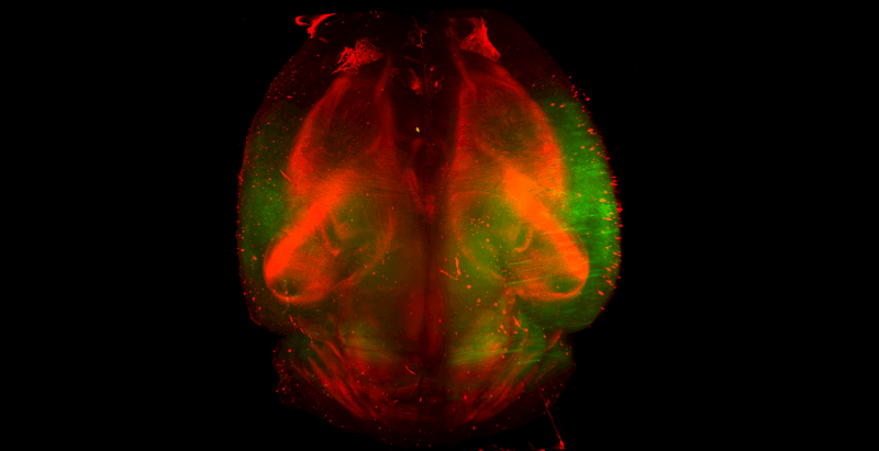
With over a dozen different tissue clearing techniques such as CLARITY, Scale, BABB, 3DISCO, SeeDB and Visikol HISTO, researchers have a wide range of tools at their disposal for the labeling and clearing of whole tissues. Each one of these techniques has their own respective advantages and disadvantages, but generally are all able to produce cleared and fluorescently labeled tissues.
Once a tissue has been cleared and labeled, the next step in the tissue processing workflow is to image the tissue and to turn your transparent tissue into a large volume of data that you can later process. While tissues can be imaged with a two-photon microscope, we are going to assume here that you are interested in multiple fluorophores and thus want to choose between light sheet and confocal microscopy.

Background of Light Sheet and Confocal Microscopes
Light sheet microscopes have been around for about a hundred years, but were not widely adopted for use in bio-imaging until the advent of fluorescent proteins, immunofluorescent labeling and other advances such as improved computing power and tissue clearing. Light sheet microscopes image tissues in 3D by passing an ultra-thin laser light sheet through a cleared tissue while imaging the laser light plane with an orthogonal objective. The objective is situated such that its focal length is in line with the laser light sheet throughout imaging where the tissue is moved through the light sheet. As the tissue is moved through the light sheet, optical Z sections are taken throughout the tissue to generate three-dimensional data sets. Generally, light sheet microscopes are most ideal for the rapid and low resolution imaging of large tissues such as a whole mouse brain.
Confocal microscopes are more complex in design than light sheet microscopes, but are significantly more prevalent in the market. Confocal microscopes image tissues in 3D by passing a laser through an imaging objective into a tissue and then collecting the resulting emitted light using a combination of dichroic mirrors and filters. Confocal microscopes are then able to generate optical Z sections from tissues by filtering out of plane light by using a pinhole confocal mechanism. Confocal microscopes are generally best suited for high resolution imaging of smaller tissues such as a 1 mm brain slice.
Considerations for Choosing Between Light Sheet and Confocal Microscopy
1. Imaging Depth: With any confocal microscope and a 10 or 20X air objective you can image 1-2 mm into a tissue before optical attenuation and refractive index mismatch cause significant image artifacts. If you want to image into a tissue past 1-2 mm with a confocal microscope, you will need high NA dipping objectives which are expensive (>$15,000) and are not compatible with several tissue clearing methods due to solvent incompatibility. Because of the optics of light sheet microscopes, they are better suited for deep tissue imaging than confocal microscopes and are designed for imaging deep into tissues. If you are working with tissues with a smaller thickness than 1-2 mm (e.g. spheroids, microtissues, organoids) always use a confocal microscope as many of these devices are setup for use with well plates for high-throughput imaging.
2. Access: Typically most universities will have either an upright or inverted confocal microscope with air objectives. It is much rarer for a university or company to have a light sheet microscope or dipping objectives. You should identify resources near you for your 3D tissue imaging as this might dictate the type of imaging that you can utilize.
3. Photobleaching: Because confocal microscopes illuminate the entire tissue thickness when acquiring an optical Z section from a single plane, they are more prone to bleaching fluorophores than light sheet microscopy. This is especially problematic if you are imaging a weakly expressed fluorophore.
4. Mounting: An important consideration for imaging a cleared tissue is how the tissue will be mounted for imaging. Tissues that have been cleared using a solvent based techniques (e.g. BABB, iDISCO, 3DISCO, uDISCO, Visikol HISTO) cannot be directly transferred to an aqueous mounting medium (e.g. VECTASHIELD) for imaging as this will cause clouding. This could potentially be problematic for imaging as your dipping objectives on your confocal or light sheet system might not be compatible with solvent based clearing techniques. Some instruments such as the La Vision Biotec Ultramicroscope II light sheet microscope are compatible with solvent based techniques, but many are not and you could potentially damage your device.
Practical Considerations and Suggestions for 3D Imaging
When considering your 3D imaging modality consider what information you NEED to collect and always ask yourself what resolution you absolutely need and if you need your tissue to be intact or if you can image several small pieces and digitally reconstruct later.
Whole brain low resolution imaging suggestion
Use a light sheet microscope if you have one and it is compatible with your tissue clearing approach or image several 1 mm sections of your tissue and piece back together digitally. If using antibody labeling it is suggested that 1 mm sections are always used if possible as it will reduce processing time by weeks compared to whole brain tissue processing.
Whole brain high resolution imaging suggestion
High resolution imaging is possible with several light sheet microscopes as well as with confocal microscopes using dipping objectives. However, high resolution imaging of whole mouse brains that are intact is incredibly challenging and becomes even more difficult if you are trying to use antibody labeling instead of fluorescent protein labeling. Researchers should also be careful with high resolution imaging of large tissues as it will take a long period of time and will potentially generate more data then you could possibly process.
