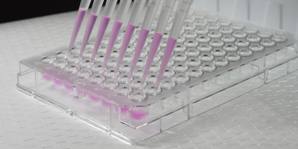
ELISA (Enzyme Linked Immunosorbent Assay) is a well-established analytical biochemical assay that is used to detect the presence of an antigen (analyte or ligand). The purpose of the assay is to quantify the amount of a specific antigen (antibodies or peptides). There are four types of ELISA’s – direct, indirect, sandwich, and competitive. In the drug discovery process the Sandwich ELISA is a great tool to infer more data from biological experiments.
Most ELISA’s are commercially available and sold as kits targeting specific antigens; generally they are sold in a 96-well format and provide all the reagents required to run the assay (including an antigen standard). When purchasing, it is important to observe the sensitivity range of your ELISA kit as many kits have sensitivities into the picogram/mL range. When performing an ELISA for the first time on a sample with uncertain concentration be sure to spare a small portion of wells for antigen concentration optimization as it is possible to oversaturate the assay, thus wasting the ELISA plate.
One of the most commonly run ELISA’s in drug discovery is the sandwich ELISA. The sandwich ELISA method is preformed in 4 steps.
- The process starts with the adherence of a capture antibody to the ELISA plate. Capture antibodies must be adhered to a cell culture plate pre-coated with a coating buffer. Common coating buffers are bicarbonate (pH 9.6) or PBS (phosphate buffered saline). These buffers increase the hydrophobicity of the plate surface, increasing interactions with amino acid side chains of the antibody. The average capture antibody is incubated overnight (2-8°C) to allow the antibody to absorb to the plate. The capture antibody is an antibody raised against the antigen of interest in order to bind specifically in the next step of the process.
- Both the sample and analytical standard are then added to the plate and allowed to incubate (with shaking). Any antigen found in the sample will adhere to the capture antibody on the plate. Standards are generally added in duplicate, and samples are generally added in triplicate.
- A detection antibody labeled with an enzyme (commonly horse radish peroxidase or alkaline phosphatase) is then added to the plate. This enzyme binds to the target antigen that was bound to the plate during sample incubation.
- Lastly, a detection antibody is introduced to the samples and elicits a colored product quantifiable using absorbance measurement. ELISA detection antibodies are usually chromogenic (molecules involved in the production of color or pigments). Most antibody kits provide detailed instructions. It is always a good idea to check the website of the vendor prior to purchasing an ELISA kit, as many of them provide the protocols needed to carry out your experiment on the product page.
This process is referred to as a “sandwich” ELISA due to the fact that the analyte being measured is bound (“sandwiched”) between two primary antibodies. It is important to completely remove any excess buffers or antibody solutions following each step within a Sandwich ELISA protocol.
An ELISA is a great endpoint for most biological experiments and one that many of Visikol’s clients incorporate into their experiments. Visikol offers an array of services such as the Blood Brain Barrier Assay, Fibrosis and Collagen Deposition, Liver Fibrosis, Hepatotoxicity, 3D Cell Culture, and many other customizable biological assays where an ELISA may be useful.
Contact us to work with our team to identify the best assay and model for your specific research needs.

