Precision-cut tissue slices (PCTS) are thin tissue slices with precise thickness generated from fresh, viable tissues through the use of a vibrating microtome (e.g. vibratome, compresstome). These slices can be cultured ex vivo and used to assess various metrics of interest to drug discovery, including pharmacological, toxicological, and physiological studies. The use of precision-cut tissue slices as an experimental model has become popular because of several major advantages over conventional 2D or 3D cell culture models. Unlike 2D or 3D cell culture models—which may lack tissue architecture, intracellular matrix, or cell heterogeneity—precision-cut tissue slices can recapitulate in vivo tissue environment because PCTS retain intact tissue architecture, extracellular matrix (ECM), cell-cell or cell-ECM interactions and heterogenous cell populations of the tissue of interest. Several studies (discussed below) have suggested that using precision-cut human tissue slices can bridge the translational gap between animal studies and human observations. Because precision-cut tissue slices are an ideal platform for pharmacology and toxicology studies, helping to overcome limitations offered by the traditional cell culture models, Visikol has established and validated service offerings using PCTS ex vivo culture model using healthy and/or diseased human or rodent tissues.
Precision-cut tissue slices from diseased human or mouse tissues can be utilized to predict toxicology, drug efficacy and therapeutic effects1-3. For example, precision-cut liver slices (PCLS) can be generated from diseased human liver with late stage liver fibrosis (e.g. F3 or F4 liver). Our data shows that precision-cut liver slices from F4 human liver can be cultured and retain viability up to at least 72 hours, and hepatocytes , immune cells, and vesicles indicative of steatosis can be maintained throughout the culture period (Fig. 1). Immunostaining of whole mount precision cut liver slices shows an extensive collagen network surrounding many vimentin-expressing hepatic stellate cells (HSC), which also very frequently express high levels of alpha smooth muscle actin indicative of HSC activation (Fig. 2).
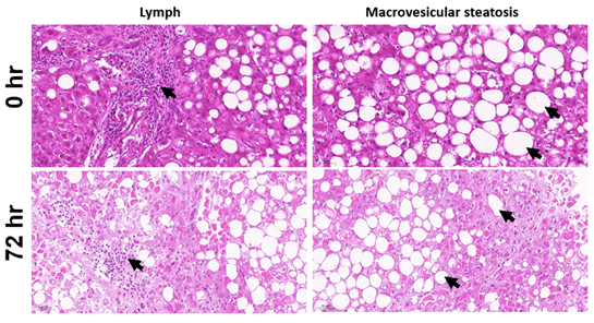
Figure 1. H&E staining showed the presence of lymphocytes and macrovesicular steatosis in human F4 precision-cut liver slices. Lymphocytes and macrovesicular can be detected at 0 hr and maintained after 72 hrs under culture conditions.

Figure 2. Structure integrity of F4 human precision-cut liver slices after 72 hours of ex vivo culture. Precision-cut liver slices of 250 µm thickness were cultured ex vivo for 72 hours. Intact precision-cut liver slices were immunostained for vimentin (red), pan-collagen (green) and alpha smooth muscle actin (ACTA2, grey).
Treatment with a specific ALK5 inhibitor in F4 human precision-cut liver slices (PCLS) reduced the expression of fibrotic genes such as metallopeptidase Inhibitor 1 (TIMP) and collagen type 1 alpha 1 (COL1A1) compared to vehicle treated PCLS (Fig. 3A). In collaboration with Melior Discovery, Visikol has also established precision-cut liver slice screening platform using carbon tetrachloride (CCl4)-induced liver fibrosis mouse model. Blocking ALK5 function in CCl4-treated mouse precision cut liver slices significantly reduced the mRNA levels of platelet derived growth factor beta (PDGFRβ) and collagen 1A1 (COL1A1; Fig. 3B). These data indicate the usefulness of precision-cut liver slices as a drug screening platform to assess compounds intended to mitigate fibrosis.
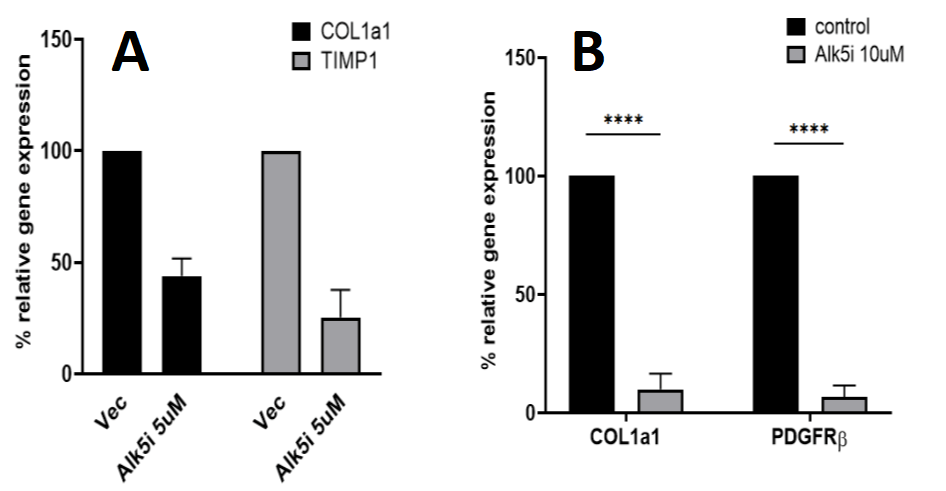
Figure 3. Gene expression analysis of precision-cut liver slices generated from F4 human liver and CCl4-induced liver fibrosis mouse model. A) Alk5 inhibition reduced the expression of COL1A1 and TIMP1 in F4 human precision cut liver slices; B) The expression of COL1A1 and PDGFRβ were significantly suppressed after Alk5 inhibition in precision-cut liver slices from CCl4-treated mice.
In addition, several studies have shown that precision-cut liver slices are an excellent ex vivo model to study viral infection and replication4, 5. Alternatively, liver fibrosis can be modeled using precision-cut liver slices ex vivo using chemicals or inflammatory cytokine cocktails6. Treatment of transforming growth factor β1 (TGFβ-1) and platelet growth factor, PDGF-BB caused fibrogenesis in precision-cut liver slices derived from both humans and rats6. Paish et al also demonstrated that anti-fibrotic compound treatment successfully attenuated ECM deposition and reduced the expression of fibrotic genes in these PCLS.
The utilization of precision-cut tissue slices is a powerful tool for drug discovery research which can yield great insights to those in pursuit of an IND for a new drug compound, or for those engaged in basic research to better understand fundamental pathways for disease. Expert utilization of precision-cut tissue slices requires the accumulation of many disciplines: 3D cell culture, pharmacology, histopathology, tissue-clearing and confocal imaging, genetics, biochemistry, and logistics. As such, rest assured that when you work with Visikol, our experts will work diligently to oversee all the details, work with you to plan an excellent experiment to your exact specifications, and give you access to unique insights for pre-clinical drug discovery. Reach out today to discuss our precision-cut liver slice and precision-cut tissue slice service offerings.
References:
- de Graaf et al., Preparation and incubation of precision-cut liver and intestinal slices for application in drug metabolism and toxicity studies. (2010) Nature Protocols (5) p1540–1551
- de Bovenkamp et al., Precision-Cut Liver Slices as a New Model to Study Toxicity-Induced Hepatic Stellate Cell Activation in a Physiologic Milieu. (2005) Toxicological Sciences (85) Issue 1, p632–638
- Westra et al., The Effect of Antifibrotic Drugs in Rat Precision-Cut Fibrotic Liver Slices. (2014) PLoS ONE 9(4): e95462
- Lagaye et al., Efficient replication of primary or culture hepatitis C virus isolates in human liver slices: a relevant ex vivo model of liver infection. (2012) Hepatology 56(3) p861–872
- Palma et al., Precision-cut liver slices: a versatile tool to advance liver research. (2019) Hepatology International (13) p51–57
- Paish et al, A Bioreactor Technology for Modeling Fibrosis in Human and Rodent Precision‐Cut Liver Slices (2019) Hepatology 70(4) p.1377-1391