
We see it as a core focus of our company to continually educate and inform our Customers and Clients on the most advanced imaging, image analysis and in vitro tools for use in drug discovery. It is for this reason that we continually generate tutorials, videos and presentations for our research community.
Visikol is a contract research services company that is focused on accelerating the drug discovery and development process through providing its clients with advanced tissue imaging and advanced cell culture services. We provide end-to-end services that include 3D tissue imaging, multiplex tissue imaging, digital pathology, high content imaging, 2D cell culture assays, 3D cell culture assays and ex vivo tissue slice assays. Visikol’s expertise lies in both transforming tissues into actionable insights as well as bridging the gap between in vitro assays and in vivo results through the use of best-in-class cell culture models.
Visikol’s Multiplex Services
At Visikol, we provide our Clients with the ability to move beyond traditional slide labeling and imaging such that they can achieve 10+ labeling from a single slide.
Visikol Expansion Ribbon Cutting
On Thursday, July 28, 2022 Visikol hosted a ribbon cutting to celebrate its expanded lab and office space.
Visikol’s Blood Brain Barrier Assay
Our Blood Brain Barrier assay is the gold-standard in vitro BBB testing platform and is now more accessible than ever.
Cytometric Analysis of Immune Cell Populations
Cytometric Analysis of Immune Cell Populations in Archived clinical FFPE Biopsies for Immuno-oncology Research.
Modern Approaches to Drug Discovery Panel
Visikol’s Dr. Tom Villani, Ph.D. and Dr. Michael Johnson, Ph.D. participated in the ‘Modern Approach to Drug Discovery Panel’ at the 2021 BICO Partnership Conference.
Tissue Clearing Webinar
In this webinar, Visikol CEO Michael Johnson, PhD provides an overview of tissue clearing and how to employ tissue clearing to image tissues in 3D using confocal or light sheet microscopy.
Aperio VERSA Visikol’s Highly Multiplexed Immunohistochemistry Approach
Through this presentation, Visikol will provide a background on various multiplex tissue imaging approaches and will describe how they can be used to answer complex research questions such as surveying the immune cell population within a tumor microenvironment or evaluating therapeutics for infectious diseases.
Case for 3D Cell Culture Models in Drug Discovery
In this video we describe how growing the Cellaria Wood breast cancer cell line in 3D and 2D generated significantly different results when used to screen small molecule drugs. Specifically, the cell line recapitulated the expression of estrogen receptor in 3D but did not express this in 2D.
Beating Primary Cardiomyocyte 3D Cell Culture
Visikol has the capability to generate a wide range of primary 3D cell culture models and in this video demonstrates its ability to generate murine derived cardiomyocyte models that are able to beat.
Visikol HISTO Brain Vasculature
Through the use of CD31 vasculature labeling combined with Visikol® HISTO™ and confocal microscopy entire vasculature networks can be mapped in 3D.
Whole Lung 3D Tissue Imaging
Complex and heterogeneous tissues such as the lung which are challenging to depict via traditional 2D histology can be easily characterized in 3D using Visikol HISTO and immunolabeling.
Virtual Reality Mouse Brain Vasculature
Data sets generated from confocal microscopy combined with tissue clearing can be visualized using virtual reality by exporting data sets as surface files.
Visikol HISTO Owl Brain Imaging
Whole brain connectomics can be executed through the use of tissue clearing combined with immunofluorescent labeling.
Visikol HISTO 3D Mouse Prostate Imaging
Biology that is challenging to depict in 2D can be easily characterized in 3D through the use of confocal microscopy combined with fluorescent labeling.
Visikol HISTO 3D Mammary Tissue
Breast cancer is heterogeneous and complex wherein important details can be missed with traditional 2-dimensional histology.
Visikol and Abcam Tissue Clearing Webinar
In this webinar, Visikol provides an overview of tissue clearing, 3D tissue imaging, 3D tissue labeling and 3D data processing.
Liver 3D Cell Culture Model Overview Tutorial
Dr. Erin Edwards provides background information on how and why liver 3D cell culture models are used in toxicity/disease modeling (e.g. NASH, NAFLD) studies.
Dr. Michael Johnson LDV Vision Summit
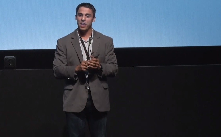
Dr. Michael Johnson discusses the future of tissue imaging at the 2018 LDV Vission Summit in NYC.
Tissue Clearing Webinar
Dr. Michael Johnson provides an overview of tissue clearing and 3D imaging.