While 3D cell culture models are being adopted in the drug discovery space for their improved in vivo relevancy, the imaging techniques used to characterize these models are highly limited. The problem with the current imaging paradigm in regard to 3D cell culture models is that due to the thickness and opacity of these models, light cannot penetrate to the center of the tissues, and so only the outer 2-3 layers of cells can be detected. This causes the dark centers often seen in images of 3D cell culture models and is highly problematic as these 2-3 cell layers are not indicative of the entirety of the model as they are most exposed to compounds, nutrients and oxygen. To address this problem, we have developed Visikol HISTO-M which is a tissue clearing technique designed specifically for 3D cell culture models and plate-based high-throughput processing. The Visikol HISTO-M technique when paired with fluorescent labeling (e.g. fluorescent protein, immunofluorescence, chemical dyes) and high content confocal microscopy allows for complete 3D cell culture model characterization and more accurate drug screening.
Why use Visikol HISTO-M?
While there are dozens of tissue clearing techniques described in the literature, there are few that have been demonstrated for use with 3D cell culture models. Additionally, most of these tissue clearing techniques are not designed for compatibility with well plates (e.g. iDISCO, BABB, uDISCO, 3DISCO) or high-throughput processing (e.g. CLARITY) and many are not compatible with immunofluorescent labeling (ScaleS4, CUBIC) or have high toxicity (e.g. ClearT). Therefore, we developed the Visikol HISTO-M tissue clearing technique to be rapid, easy-to-use, plate compatibility, fluorescent protein compatible and immunofluorescence compatible.
Buy Visikol HISTO-M Products
To get started with Visikol HISTO-M you can purchase the Visikol HISTO-M Starter Kit from the Visikol store or you can purchase the Visikol HISTO-M reagent and then make the required buffer reagents yourself.
How to get started?
- Purchase the Visikol HISTO-M reagent from the Visikol store.
- Purchase or make the Visikol HISTO buffer reagents.
- Read through the Visikol HISTO-M guidebook to generate a customized protocol.
- Follow the getting started guide.
Benefits of Visikol HISTO
- Rapid tissue clearing
- Easy-to-use
- No special equipment required
- Compatible with IF, FP and other fluorescent labels
- Well plate and automation compatible
- Reversible for follow up 2D H&E/IHC
Complete model characterization
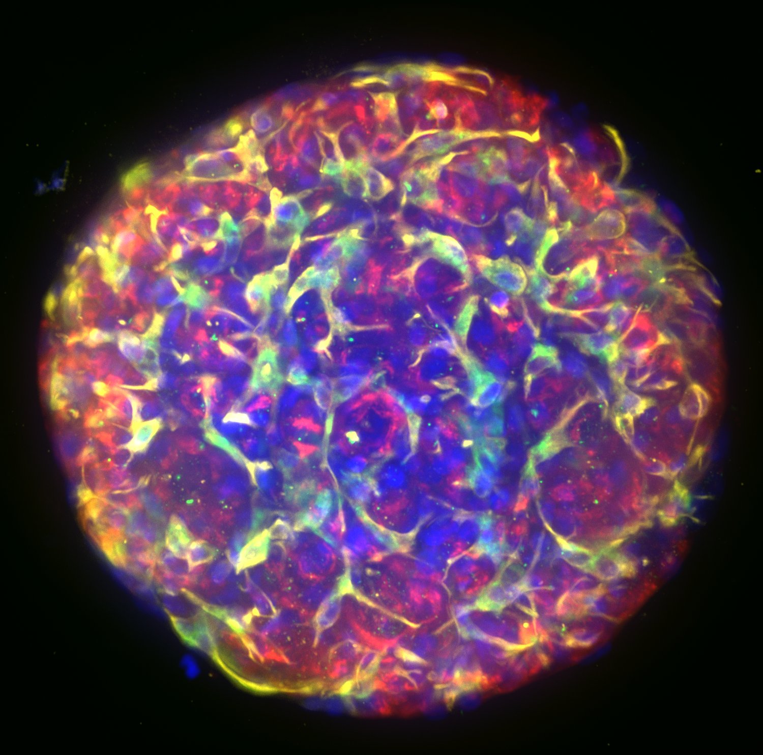
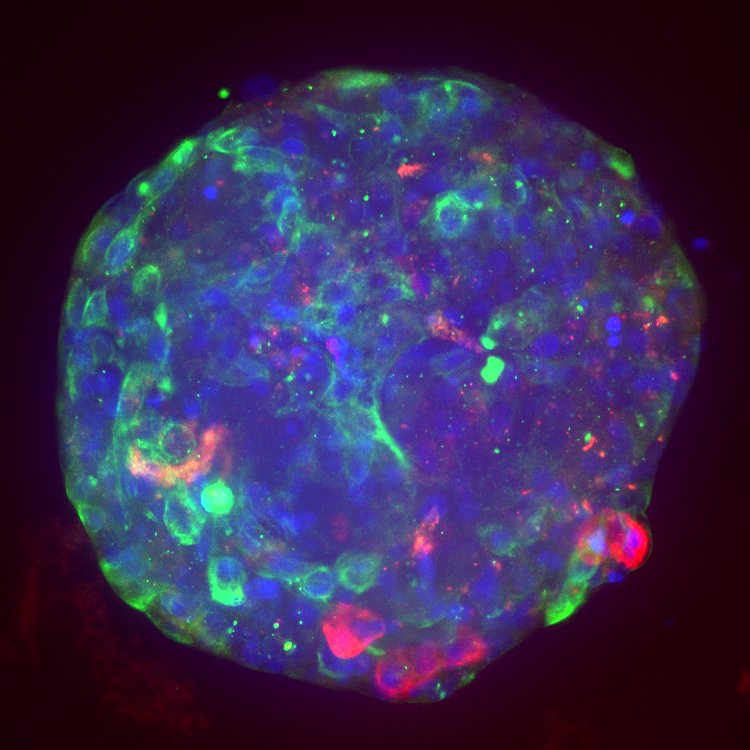
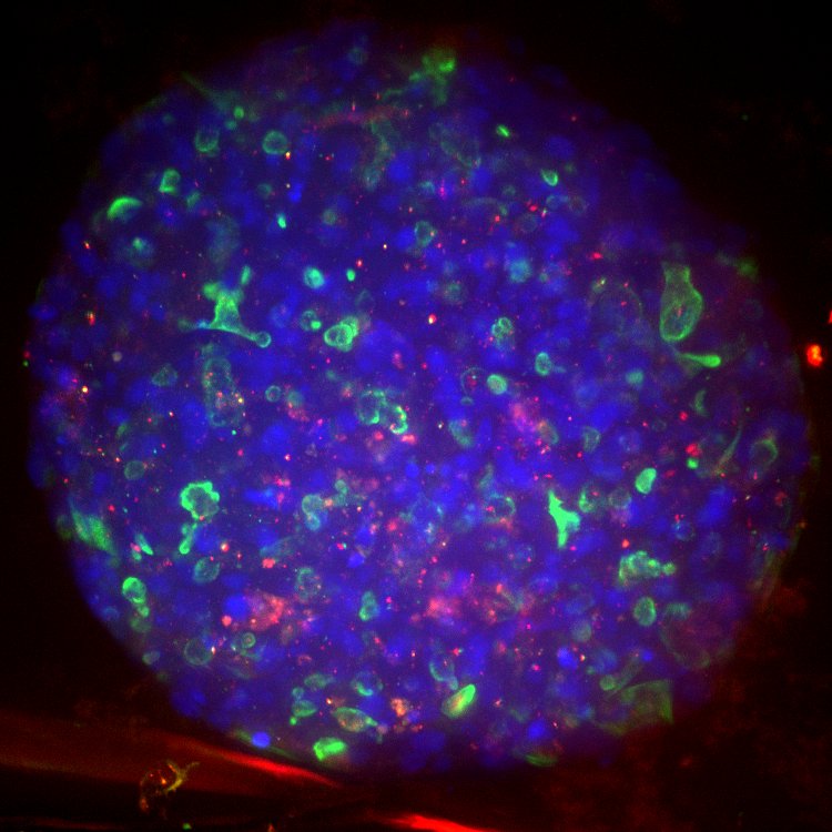
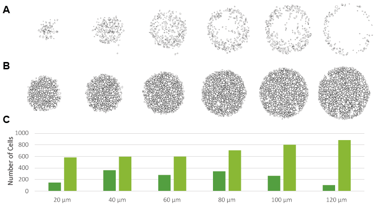
The use of Visikol HISTO-M enables researchers to visualize 3D cell culture models in their entirety through the use of confocal microscopy which results in a 3-4 increase in the number of cells detected. The technique is compatible with flat bottom plates as well as ultra low attachment U-bottom plates.
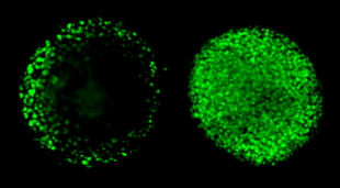
Before and after clearing.
Below it is clearly shown tissue clearing combined with confocal microscopy allows for a 3-4 increase in cells detected (C) when compared a 3D cell culture model cleared with Visikol HISTO-M (B) and a 3D cell culture model imaged in PBS without clearing.
Increased dose response sensitivity
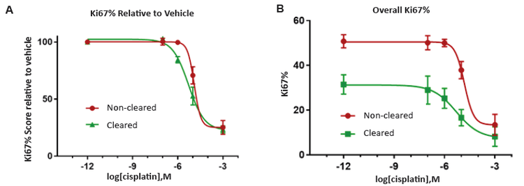
The use of Visikol HISTO-M allows for a significant increase in dose response sensitivity (left) as the technique allows for the entire population of cells within a 3D cell culture model to be characterized. Furthermore, due to interrogation of the entire cell population, assessment of drug effect is more accurate. Without clearing, only the exterior cells are surveyed, and as such, measurements of cell proliferation are overestimated, since the outer layers of cells show more proliferation than the interior cells.
