In recent decades, the increase in computing power and the development of automated microscopes and high-performance slide scanners have revolutionized the field of bioimaging. With the ability to process vast amounts of data, researchers are now able to glean new insights and drive innovation in their respective fields. However, the complexity of analyzing such large datasets requires expertise in both biology and computer science. At Visikol, we provide quantitative image and data analysis services for high content and whole slide image datasets, either as a standalone service or in conjunction with our assay and tissue imaging services. With our cutting-edge technology and experienced team, we routinely process terabytes of images per day, ensuring that no project is too large or small. Let us help you unlock the potential of your biological imaging data and take your research to the next level.
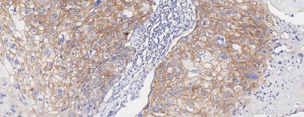
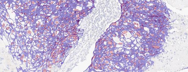
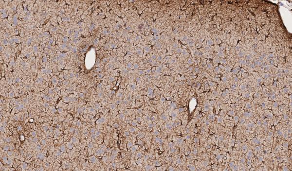
Quantification of positive cells for immunohistochemistry
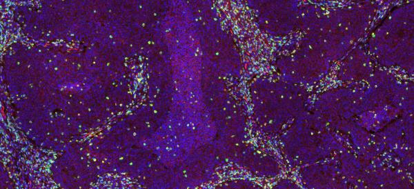
Analysis of immune cell population within and outside of tumor for immuno-oncology
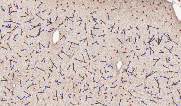
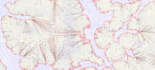
Analysis of spatial relationships between cell types
Image Analysis Endpoints
Available on: