
NEW BRUNSWICK, N.J. (PRWEB) APRIL 15, 2018
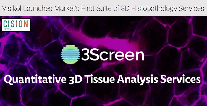
For as little as $500 per tissue sample, Visikol is now offering 3D tissue labeling and imaging services so that all researchers can easily and quickly acquire 3D information from their tissues. This service offering is part of Visikol’s 3Screen™ services and is as easy as sending a tissue sample and then receiving a USB drive with over 1,000 images.
Since the introduction of the microscope, researchers and clinicians have characterized tissues using ultra-thin slices that are placed on glass microscope slides and selectively stained. While staining technologies (e.g. antibody labeling, FISH) have advanced dramatically since the introduction of the microscope, the practice of slicing whole tissues into ultra-thin sections has been largely unchanged and forms the basis of many life science fields today including clinical diagnosis.
However, many tissues are complex and heterogenous where a few ultra-thin sections could miss histologically important features such as a micrometastases between slices or incompletely characterize a tissue. “The current histology paradigm typically relies upon less than one percent of the data within a tissue to evaluate the entire tissue and this evaluation is traditionally conducted in a qualitative human-driven approach,” describes Visikol Director of Research Dr. Graeme Gardner. The limitation of this current paradigm is that very little data is generated from tissues and this data has always been in a non-digital format that is left up to subjective interpretation.
To address this current limitation, Visikol has launched its 3Screen whole mount tissue imaging services which enable every researcher to quickly transition from acquiring a few images from a tissue to hundreds of images throughout the depth of a tissue without the need for serial sectioning. Visikol is offering this service for as little as $500 per tissue with a two week turn-around time. “The ability to now add spatial information to histology means that researchers can quantitatively study complex features such as mapping the growth of blood vessels in a tumor or the diffusion of nanoparticles through a tissue. Dramatically increasing the amount of data acquired from a single tissue also allows researchers to apply machine learning and artificial intelligence algorithms to these data sets for advanced analysis and acquiring additional insights not possible with traditional histopathology,” explains Visikol Chief Science Officer Dr. Thomas Villani.
Visikol’s whole mount tissue imaging services combine the companies patented Visikol® HISTO™ tissue clearing technique with fluorescent immunolabeling and high throughput confocal microscopy. These services allow researchers to optimize their labeling and imaging parameters based upon what is required for their specific research question.
“The focus of our company has always been on transforming tissues into three-dimensional data sets that can be quantitatively analyzed to provide researchers with actionable insights. With this new service offering, we are providing researchers who do not have the resources or time to adopt tissue clearing in their lab the ability to generate vast amount of feature-rich histological data from their tissues,” explains Visikol Chief Executive Officer Dr. Michael Johnson.
To get started with 3D tissue imaging, the process is as simple as sending along a piece of tissue and waiting two weeks until a USB containing all of the relevant data is sent along. The Visikol team has optimized a suite of antibody labels for use with 3D tissue imaging and can also optimize new labels for use in 3D to suit a specific researcher’s need.
Interested in learning more about our advanced imaging and drug discovery services? Download our Services Catalog
