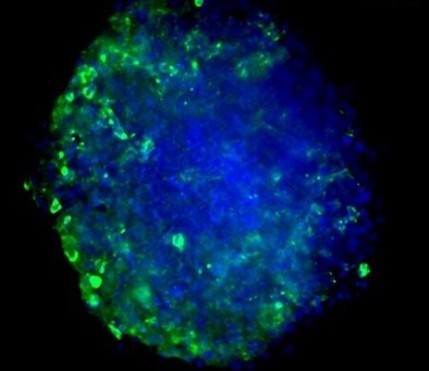
 In vitro immuno-oncology models are used to study the interaction of therapeutics with immune cells in a 2D or 3D assay format. These models are used to assess the effect of compounds and therapeutic antibodies on immune cell infiltration or to screen immune cell populations for use in immunotherapy. The 3D cell culture models are more biologically relevant and allow for proper assessment of immunotherapies designed to promote the immune cell response to tumor cells. Visikol’s in vitro immuno-oncology models and high content imaging technology can address limitations by providing a method for improving in vitro studies used in drug discovery and development. In this blog post, we will explore how Visikol’s in vitro immuno-oncology models can be used for high content imaging, allowing for more accurate and reliable data for drug efficacy and safety testing. We will also discuss how Visikol’s 3D cell culture models are more biologically relevant and how their high content imaging technology can help researchers and pharmaceutical companies maximize the potential of their immuno-oncology models.
More on High Content Imaging
High content imaging technology is a method of in vitro assay services that allows for the assessment of multiple endpoints simultaneously at a cellular resolution. It utilizes imaging-based endpoints to examine the specific effect of compound treatments on specific sub-populations of cells, as well as providing access to more complex measurements than can be accomplished with traditional assay formats. The data generated from high content imaging is processed using purpose-built image processing pipelines to obtain quantitative data, extracting cell counts, morphological features, colocalization of labels, and computing statistical comparisons between groups simultaneously.
High content imaging technology allows for the assessment of multiple endpoints simultaneously at a cellular resolution, providing richer datasets than traditional assays. This approach enables the interrogation and quantitation of cellular response of disease models to treatments, stimuli, or alterations in protein expression. The use of imaging-based endpoints allows for examination of the specific effect of compound treatments on specific sub-populations of cells, as well as providing access to more complex measurements than can be accomplished with traditional assay formats. Combining these data with machine-learning and informatics expertise, comprehensive reports can be generated that distill the data to its essential components, giving actionable insights about the experiment.
Visikol’s in vitro Immuno-Oncology Models
Visikol’s in vitro immuno-oncology models and high content imaging technology can address limitations by providing a method for improving in vitro studies used in drug discovery and development. They offer tissue analysis services for immuno-oncology research, which aims to understand how the immune system interacts with the tumor microenvironment and modulate the immune response to reduce tumor growth. They employ multiplex immunofluorescence to simultaneously interrogate multiple cell populations and use automated image processing algorithms to assess the variety and distribution of immune cells within and around tumor tissue. They also offer High Content Screening services to clients utilizing 3D cell models and traditional cell-based assays. They provide a wide range of validated in vitro screening services, rapid custom assay development, and end-to-end compound screening services.
3D Cell Culture Models
3D cell culture models are a method of growing cells that better replicates the complex characteristics of the in vivo microenvironment, such as diffusion gradients and receptor expression. Traditional 2D cell culture is limited in its ability to replicate these characteristics. The technique in which cells are cultured (2D vs. 3D) can substantially alter the drug’s effect on the cells. 3D cell culture models offer a more natural, tissue-mimicking method of cell growth for drug discovery applications.
Visikol’s expertise in 3D cell culture models and high content imaging technology allows for the assessment of multiple endpoints simultaneously at a cellular resolution. This provides richer datasets than traditional assays, enabling the interrogation and quantitation of cellular response of disease models to treatments, stimuli, or alterations in protein expression. The use of imaging-based endpoints allows for examination of the specific effect of compound treatments on specific sub-populations of cells, as well as providing access to more complex measurements than can be accomplished with traditional assay formats.
Visikol’s in vitro immuno-oncology models and high content imaging technology can provide a method for improving in vitro studies used in drug discovery and development. Their 3D cell culture models are more biologically relevant and allow for a proper assessment of immunotherapies designed to promote the immune cell response to tumor cells. Their high content imaging technology can help researchers and pharmaceutical companies maximize the potential of their immuno-oncology models by providing a method for assessing multiple endpoints simultaneously at a cellular resolution. By utilizing these technologies, researchers can obtain more accurate and reliable data for drug efficacy and safety testing, ultimately leading to the development of more effective cancer treatments.
If you’re interested in learning more, please reach out to a member of our team today!
