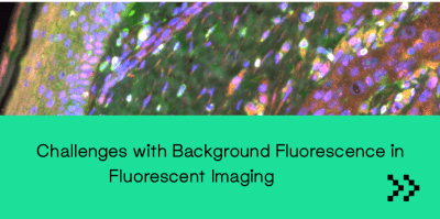
Those who have done fluorescence microscopy and imaging are aware that autofluorescence can make imaging and data interpretation difficult. What is most often referred to as “autofluorescence” is “natural fluorescence” or “fixative-induced fluorescence”. Usually, the emission spectra of natural fluorescence and fixation-induced fluorescence are very broad compared to the spectra of the dyes, probes, and biomarkers. This makes it difficult to separate wanted from unwanted fluorescence by traditional filtering methods. The best ways to address the issue of autofluorescence are by trying to filter it out during image acquisition, which is difficult due to its broad emission spectrum of autofluorescence, or by chemically removing it. Natural fluorescence is usually due to substances like flavins, porphyrins, chlorophyll, etc. They may persist, or even become redistributed, during the fixation and dehydration and may be present in frozen sections that have passed through various aqueous reagents.
Lipofuscin
A native autofluorescent material that persists even after formalin fixation. Lipofuscins are brown in color; its excitation spectrum is UV-blue and emission spectrum is green-yellow-orange. Lipofuscins are prominent in large neurons, glial cells, cardiac muscle cells and stains positive for proteins, carbohydrates, and lipids. It’s a product of old red blood cells and are strongly fluorescent under any excitation ranging from 360nm to 647 nm. It may be quenched with either CuSO4 or Sudan Black.
Sudan Black treatment protocol: 1. 0.3% Sudan Black (w/v) in 70% EtOH (v/v) stirred in the dark for 2 hours 2. Apply to slide for 10 minutes after the secondary antibody application. 3. Rinse quickly with PBS 8 times and mount 4. For FITC and Alexa 594 this does not reduce the emission signal noticeably.
Elastin
Contains several fluorophores; this is a similar fluorophore to that found in collagen. All blood vessels on the arterial side of the circulation muscle cells have it. Elastin is intensely fluorescent over a range of excitation wavelengths. The autofluorescence coming from the elastin fibers, can be quenched by incubating labelled sections in 0.5% Pontamine sky blue 5BX in buffer or saline for 10 minutes, followed by a 5-minute wash before mounting. This shifts the usual green emission of elastin into a red emission. This works well in lung tissue using fluorescein labelled probes.
Aldehyde
Fixatives react with amines and proteins to generate fluorescent products.The simplest way to stop aldehyde-induced fluorescence is to use a fixative that does not contain an aldehyde. Eg: Methanol Carnoy fixation. Glutaraldehyde exists as low polymers, it reacts with and cross-links protein molecules, this results in free aldehyde groups. These tissue-bound free aldehyde groups will combine covalently with any amino group offered to them, including terminal and sidechain (lysine) amino groups of proteins being used as histochemical reagents – eg: antibodies, lectins and enzymes, resulting in nonspecific binding of antibodies to tissue. This free aldehyde problem can be solved by aldehyde blocking: it’s done by reducing the -CHO groups to -OH with sodium borohydride or by feeding them bland amino groups (e.g., glycine). Another way to avoid aldehyde induced autofluorescence is by giving pre-bleach treatment. A lightbox containing four fluorescent-light tubes, each with different emission peaks (UV to 633 nm) is used to bleach the aldehyde-induced fluorescence. (Neumann and Gabel 2002).
Interested in speaking to a member of our team regarding autofluorescence? Reach out today.
