Indica Labs presented a poster titled ‘Cytometric Analysis of Immune Cell Populations in Clinical Tumor Biopsy Tissue Microarrays for Immuno-oncology’ done in partnership with Visikol at AACR on April 13, 2022.
Introduction
Cancer is a deadly complex disease that affects millions around the world each year. Variability in disease etiology leads to challenges in selecting treatments that will maximize the patient’s quality of life and overall survival. It is essential for health care providers to know the extent of a patient’s cancer to better prescribe a treatment to improve prognosis. However, current methods of patient diagnosis are qualitative and/or semi-quantitative and may not include the impact of the tumor micro-environment on patient treatment and outcome. Therefore, we have developed a method utilizing 12-plex fluorescence imaging, quantitative image analysis, as well as cytometric analysis of immune cell populations to better categorize patients and determine the best treatment strategy for the individual patient.
Materials and Methods
Multiplex labeling was completed on human breast cancer tissue microarray (TMA) (XBra089-01) using traditional immunofluorescence labeling techniques. The TMA was rehydrated through decreasing ethanol concentrations, followed by antigen retrieval in citrate buffer. Labeling of the TMA was completed in 4 panels with a control nuclear stain DAPI, designed to reduce cross-reactivity and promote labeling sensitivity. Removal of antibody labeling between panels was completed using Visikol Inc’s proprietary stripping reagent EasyPlexTM. Imaging was completed at 40x on the Leica Versa 8 slide scanner.
Once the fluorescence imaging was complete, HALO v3.3 was used for the analysis by first using the HALO TMA module to tile the cores into individual image sets. The individual cores were analyzed using the HALO Highplex module v4.0.4, to perform cell segmentation and marker colocalization. Subsequently, the HALO Spatial Analysis module was utilized to perform a proximity analysis between different cell subtypes. After exporting this data from HALO as a .csv file, well-established python libraries (Pandas, SciPy, and Scikit-Learn), were used to analyze the data.
Quantitative measurement data were normalized and standardized using the standard scaling technique resulting in mean-centered values representing the number of standard deviations from the mean. To select a subset of features based on their contribution to the variation between groups, feature selection was performed using the F-test. To visualize patient population-wide variation and clustering from the selected feature subsets, principal component analysis (PCA) was conducted. Clustering of samples and measurements was conducted using hierarchical agglomerative clustering of the Euclidean distance matrix from selected feature subsets.
Results and Discussion
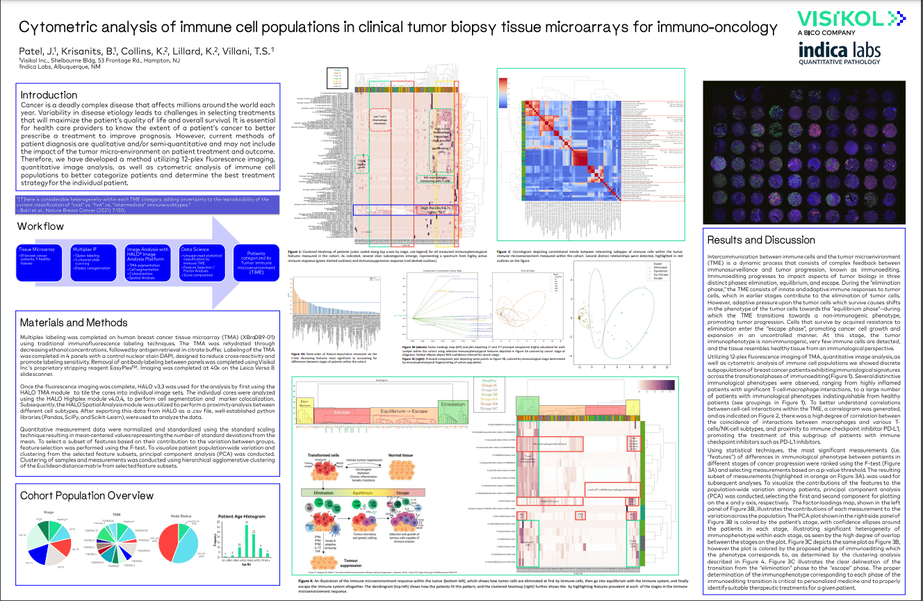
Intercommunication between immune cells and the tumor microenvironment (TME) is a dynamic process that consists of complex feedback between immunosurveillance and tumor progression, known as immunoediting. Immunoediting progresses to impact aspects of tumor biology in three distinct phases: elimination, equilibrium, and escape. During the “elimination phase,” the TME consists of innate and adaptive immune responses to tumor cells, which in earlier stages contribute to the elimination of tumor cells. However, adaptive pressure upon the tumor cells which survive causes shifts in the phenotype of the tumor cells towards the “equilibrium phase”—during which the TME transitions towards a non-immunogenic phenotype, promoting tumor progression. Cells that survive by acquired resistance to elimination enter the “escape phase”, promoting cancer cell growth and expansion in an uncontrolled manner. At this stage, the tumor immunophenotype is non-immunogenic, very few immune cells are detected, and the tissue resembles healthy tissue from an immunological perspective.
Utilizing 12-plex fluorescence imaging of TMA, quantitative image analysis, as well as cytometric analysis of immune cell populations we showed discrete subpopulations of breast cancer patients exhibiting immunological signatures across the transitional phases of immunoediting. Several distinctive immunological phenotypes were observed, ranging from highly inflamed patients with significant T-cell-macrophage interactions, to a large number of patients with immunological phenotypes indistinguishable from healthy patients. To better understand correlations between cell-cell interactions within the TME, a correlogram was generated, and there was a high degree of correlation between the coincidence of interactions between macrophages and various T-cells/NK-cell subtypes, and proximity to immune checkpoint inhibitor PD-L1, promoting the treatment of this subgroup of patients with immune checkpoint inhibitors such as PD-L1 inhibitors.
Using statistical techniques, the most significant measurements (i.e. “features”) of differences in immunological phenotype between patients in different stages of cancer progression were ranked using the F-test and selecting measurements based on a p-value threshold. The resulting subset of measurements was used for subsequent analyses. To visualize the contributions of the features to the population-wide variation among patients, principal component analysis (PCA) was conducted, selecting the first and second component for plotting on the x and y axis, respectively. The proper determination of the immunophenotype corresponding to each phase of the immunoediting transition is critical to personalized medicine and to properly identify suitable therapeutic treatments for a given patient.