
 Assessing the distribution of biologicals, such as antibody therapeutics, in solid tumors is a complex process that presents several challenges. The transport of biologicals into tissues involves a combination of factors, including diffusion, convection, and affinity to target antigens within the interstices and cell surfaces. In healthy tissues, extracellular fluid flows steadily from the capillaries to the lymphatic vessels, driving transport of macromolecules throughout the tissue. However, in solid tumors, functional lymphatic vessels are rare, leading to an increase in hydrostatic pressure, reducing the propensity of the convection gradient to drive macromolecules into the tumor.
Assessing the distribution of biologicals in tumor tissue, especially antibody drugs, has been recognized as a major issue for immunotherapy. Traditional in vitro models have been limited in their ability to predict in vivo efficacy and toxicity due to their inability to replicate the complex in vivo microenvironment. Animal studies are typically required to assess the distribution of biologicals, which is a costly and time-consuming process.
Antibody Pharmacokinetics Assay
Visikol’s in vitro Antibody Pharmacokinetics Assay is a cost-effective and time-saving alternative to animal studies for drug development. The assay measures the distribution of biologicals, including antibody drugs, into solid tumor models using tumor spheroid models and high content confocal imaging to assess the distance and quantity of antibody penetration into tumor spheroids. The assay can screen the relative differences in distribution of various biologicals, including antibody drug conjugates, antibody fragments, alternate domain antibodies, affibodies, and nanobodies. The assay can measure distribution curves, velocity curves, TD50 values, and concentration-time curves. This assay helps overcome the challenges of assessing antibody distribution in solid tumors by providing a method for screening the relative differences in distribution of these molecules in vitro, without the need for animal studies.
The use of tumor spheroid models for assessing the distribution of biologicals, including antibody drugs, into solid tumor models has become increasingly popular in recent years. Tumor spheroids are three-dimensional cell culture models that mimic the in vivo microenvironment of solid tumors more closely than traditional two-dimensional cell culture models. They offer an excellent format for confirming results of in vitro screening using 2D cells, and for screening hits and lead compounds for potential toxic liabilities.
Antibody Pharmacokinetics Assay Protocol
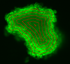
The protocol for the in vitro Antibody Pharmacokinetics Assay involves generating free-floating tumor spheroids, treating them with antibody therapeutics at various concentrations, fixing them at specified time points, labeling the antibody with fluorescent secondary to label therapeutic antibody, applying tissue clearing to render tumor spheroids transparent, high content confocal imaging, and analysis of resultant images to assess distance and quantity of antibody penetration into tumor spheroid. The test article concentration involves an 8-point assay and a single point assay, with custom concentrations available. Time points include 0.5 hr, 1 hr, 2 hr, 4 hr, 8 hr, 24 hr, 48 hr, with custom time points available. Three replicates per concentration are required, with 0.5% DMSO as the vehicle control and anti-β-integrin as the positive control. Antibody concentration is measured as a mean of pixel intensity in concentric bands of decreasing radius from the edge of the tumor spheroid into the center. The resultant data includes distribution curves, velocity curves, TD50 values, and concentration-time curves (measured as AUC).
The findings from in vitro models can be translated to in vivo models to some extent. The research on assessing antibody distribution in solid tumors using tumor spheroid models and high content confocal imaging could have potential implications for cancer treatment. It offers a cost-effective and time-saving alternative to animal studies for drug development and could help screen the relative differences in distribution of various biologicals, including antibody drug conjugates, antibody fragments, alternate domain antibodies, affibodies, and nanobodies. This could lead to the development of more effective immunotherapies for cancer treatment.
This assay helps overcome the challenges of assessing antibody distribution in solid tumors by providing a method for screening the relative differences in distribution of these molecules in vitro, without the need for animal studies. To learn more about the Antibody Pharmacokinetics Assay, please reach out to a member of our team today.