
Precision-Cut Liver Slice Study Formats

There are three possible approaches to the use of precision-cut liver slices for the assessment of drug effect ex vivo. Firstly, normal and “healthy” human donor tissue may be obtained, and a disease state induced ex vivo by chemical/biochemical stimulation, followed by treatment with test articles to evaluate potential amelioration of disease. Secondly, diseased human tissue may obtained by careful screening of donors, followed by stimulation of precision-cut liver slices to evaluate test articles for therapeutic effects. And thirdly, using animal tissue, disease state may be induced in vivo using chemical/biochemical stimulation, or by using genetically engineered models of disease, and compounds of interest may be interrogated ex vivo using precision-cut liver slices from the model animals.
Normal Human Tissue, Ex Vivo Disease Induction
Using our network of providers, we obtain F0-F1 human liver tissue. Upon receipt, tissue is prepared, precision-cut, and pre-cultured for 5 hours. Fibrosis is induced with TGF-β and/or PDGF-ββ (other stimulation conditions available on request).
Advantages:
- Relatively easy to acquire normal liver tissue
- Can induce disease state to relatively similar extent by using same induction protocol each experiment
Disadvantages:
- Disease state that is induced may exhibit non-physiological architecture or pathway


- Fluorescence microscopy depicting nuclei (DAPI, blue) and collagen (red) labeling in vehicle control, free fatty acid (FFA) stimulated, and FFA stimulated-ALK5i treated PCLS (left).
- Bar graph depicts collagen area as percent of total tissue area for different treatments.
- FFA stimulation induced collagen deposition
- ALK5 inhibitor treatment reduced FFA-induced collagen deposition
Diseased Human Liver Tissue
We obtain F3 or F4 liver tissue from donors to prepare precision-cut liver slices. Highly specific selection criteria may be applied in screening process (e.g. alcoholic vs. non-alcoholic, BMI, etc.).
Advantages:
- Most physiologically relevant disease state
- Captures native, pathophysiologically-relevant disease progression
Disadvantages:
- Somewhat longer timeframe to attain diseased liver tissues (usually acquired as explants rather than non-transplantable donor tissue)
- Disease state variability from donor to donor

Photograph of F3 human liver, showing distinct yellow fatty deposits (left). H&E image of liver shown on right.
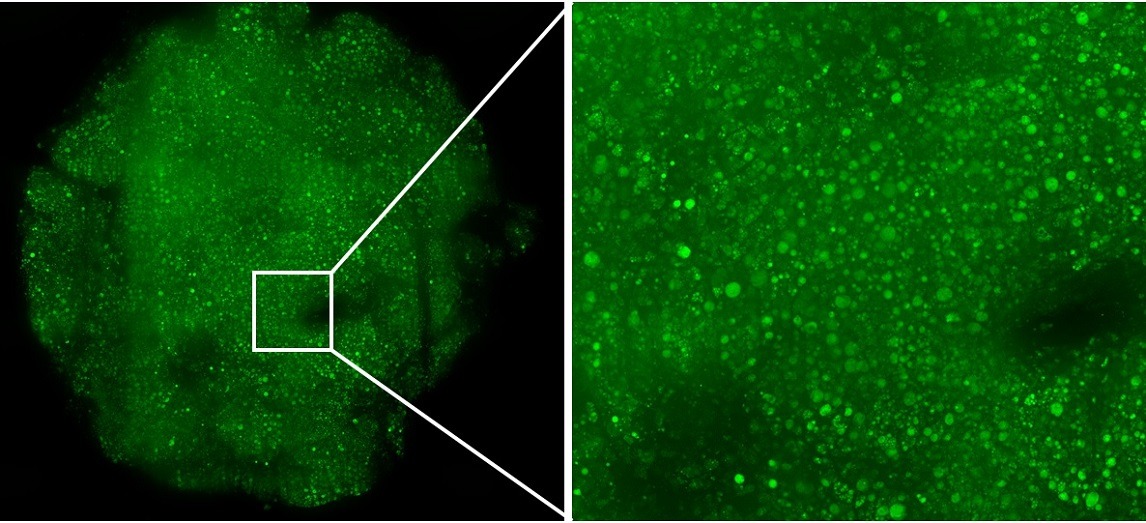
Fluorescent image of lipid droplet staining (LipidTox) illustrating lipid droplet accumulation in diseased human liver.
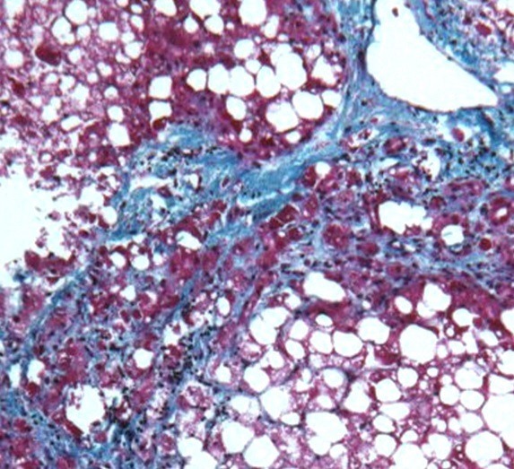
Histological section of diseased F3 human liver stained with Masson’s trichrome illustrating large areas of collagen deposits consistent with fibrosis.

Diseased F3 human liver, image taken using confocal fluorescent microscopy.

Representative relative gene expression obtained via RT-qPCR of RNA extracted from precision-cut liver slices treated with ALK5 inhibitor and vehicle control.

Results of IL-6 ELISA assay from supernatant media from precision-cut liver slices treated with Alk5 inhibitor and vehicle control.
Mouse Model with CCl4-Induced Liver Fibrosis
In collaboration with Melior Discovery, we have developed and validated the use of precision-cut livers slices derived from mouse models of liver fibrosis via 2-week induction with CCl4 for the assay of compounds to ameliorate fibrosis. Utilizing precision-cut liver slices derived from mouse models allows for predictive screening of compounds at an earlier stage, to reduce the number of animals required in later stage development.
Advantages:
- Highly reproducible study conditions due to use of animal models and specific disease induction
- Allows for genetically modified models and disease stimulation not possible in human tissue
- Reduces number of animals required in later stage studies
Disadvantages:
- Animal tissue instead of human tissue
- Simulation of disease rather than natural disease progression

Precision-cut liver slices prepared from prevalidated 2 week CCl4 mouse model (Melior Discovery). No pre-culture prior to treatment. Treated for 72h with ALK5 inhibitor or imatinib. Imatinib and ALK5 inhibitor treatment reduces collagen and PDGFRβ gene expression. Imatinib reduces PDGFRβ and αSMA expression by immunofluorescence.

