–

–

In launching the Visikol® HISTO™ technique and working with hundreds of research groups from around the world, we have heard the same feedback again and again:
“I couldn’t get tissue clearing to work in my lab.”
We have spent countless hours digging into this problem and trying to understand the disconnect between publications citing the use of a multitude of tissue clearing techniques and researchers struggling to adopt them. What we have found out is that while all tissue clearing techniques have their advantages and disadvantages, they all generally work and the problem with tissue clearing is not the techniques but instead how they are implemented.
Tissue clearing is the confluence of many complex disciplines (tissue labeling, tissue clearing, 3D microscopy, data processing) and the major challenge that underlies most of the unsuccessful applications of tissue clearing is that every application requires its own protocol and optimization approach. There are many practical considerations for 3D tissue imaging such as tissue thickness and optical limitations that need to be considered prior to implementing a tissue clearing protocol. For example, immunolabeling and imaging a whole mouse brain will require a very different protocol than what is used for clearing and imaging a neuronal organoid.
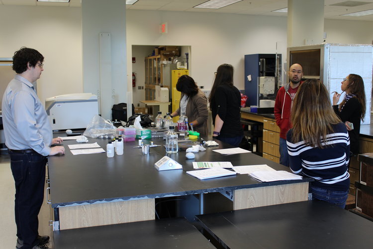
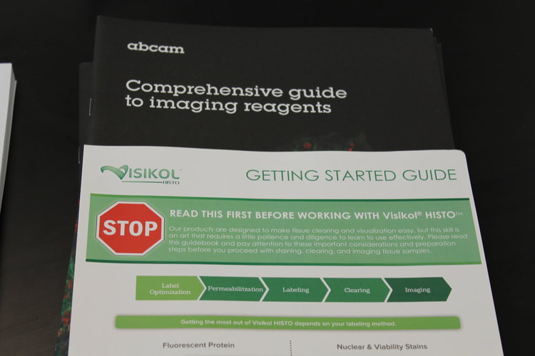
To address this problem, we partnered with over 400 research labs from around the world and launched our Protocol Guidebook that enables any researcher to customize a Visikol HISTO protocol specifically for their research question. Additionally, in 2018 we have begun to host tissue clearing workshops where our team travels to research labs and gives hands-on demos on tissue clearing, 3D microscopy, whole mount labeling and data processing.
If you are interested in hosting a tissue clearing workshop at your university email us.
