
Over the holidays, the Visikol team had the opportunity to update the highly popular Tissue Clearing Ebook. If you are interested in learning more about tissue clearing in general or how to apply tissue clearing to your research, please take a look at this comprehensive guide. The Tissue Clearing Ebook provides 3D imaging guidance, labeling guidance and all sorts of background on tissue clearing and the important considerations when adopting and applying it.
If you want to learn even more about tissue clearing, check out the great additional resources below:
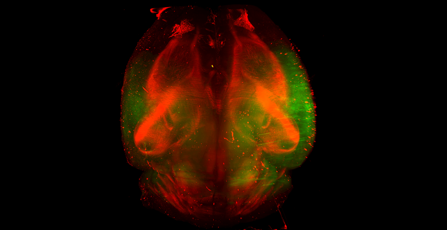
Looking Back on Tissue Clearing-Almost a Decade Later
Interested in learning more about tissue clearing? The link above covers everything from how it started (spoiler: it didn’t start in 2013 at Stanford), how it’s going, and the leaders at Visikol think the future of tissue clearing will be.
Visikol Tissue Clearing Comparison
Have you found yourself wondering ‘Which Tissue Clearing Technique is Best for Me?’ This question unfortunately does not have an easy answer. and will depend upon your specific research question. Each tissue clearing technique has advantages and no technique is best for all applications. Click the link above for a review of the pros and cons of Solvent clearing techniques, Aqueous hyper-hydrating clearing techniques, and Hydrogel embedding techniques are discussed.
Whole Tissue Imaging Guidance
In order to achieve 3D volume imaging, you need to use a confocal microscope, a light sheet microscope, or a single/multiphoton microscope. There are several major considerations to keep in mind when imaging your tissues in 3D to ensure that you achieve the best results while spending the least amount of time optimizing your protocol.
3D Cell Culture Imaging Guidance
Interested in learning more about 3D cell culture imaging? Click the link above to watch Dr. Michael Johnson, Ph.D. present on 3D cell culture characterization at the NIH.
Light Sheet Microscopy Overview
In this video Dr. Michael Johnson, Ph.D. gives an introduction to light sheet microscopes and how they can be used to image cleared and labeled tissues in 3D. Dr. Johnson gives an overview of how light sheet microscopes operate and the advantages and disadvantages with some common systems.
Confocal Microscopy Overview
In this video Dr. Michael Johnson gives a brief overview of confocal microscopy and how it is used for the 3D visualization of cleared and fluorescently labeled tissues.
Visikol HISTO Getting Started Guide
In this video Visikol CEO Dr. Michael Johnson, Ph.D. describes how to implement the Visikol HISTO tissue clearing approach into your research workflow. He discusses how best to adopt tissue clearing, labeling considerations and imaging considerations as well.
Interested in learning more about tissue clearing for your project? Contact us today!