The ability to detect cancer and changes within cancer cells is an asset in the clinic and research lab. Whether changes detected be the location, size, type, rate of growth, metastatic progression and more. Having the ability to center in on these cells and make a precise diagnosis can be the determining factor in patient outcome, treatment, and quality of life. Amongst the numerous staining and labeling techniques for cancer cells, Visikol performs Hematoxylin and Eosin (H&E) stains on various types of cancer samples and species, to help recognize the morphologic changes that occur, patterns, shapes, and cellular structures.

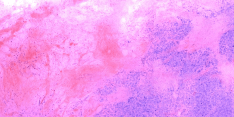
Virtual H&E image of tissue derived from an optical section taken from a thick piece of FFPE breast cancer lumpectomy tissue.
What is H&E Staining?
H&E staining is a structural stain that utilizes two main chemicals to help visualize these structures, hematoxylin, and eosin. Hematoxylin is not technically a dye and is used in combination with compounds such as metal cations (ex. aluminum). The combination of hematoxylin and cations creates a positive charge which interacts with negatively charged components of the tissue, such as nucleic acids within the nucleus, resulting in a purplish-blue color stain within the cell nuclei. Eosin is an acidic dye and is negatively charged, promoting its interaction with positive charged structures within the tissue, such as proteins in the cytoplasm, resulting in pink coloring observed in the cell cytoplasm. The combination of these two stains will give you the general layout of your cells as well as the distribution and structure of them. A major reason this stain is used is the ability to perform the test quickly with great accuracy, and is why at Visikol, it is one of our most used staining methods.
H&E Staining Process
Following tissue processing, embedding, and sectioning, tissue slices are de-waxed in Sub-X (alternatively, Clear-rite 3) and rehydrated in decreased concentrations of EtOH, followed by tap water. Once rehydrated, slides are incubated in Gill’s Hematoxylin, rinsed, and washed in tap water until all the excess Hematoxylin is removed. Next is the differentiation step, utilized to remove non-specific background staining while improving contrast (ex. 0.3% Acid Ethanol). After the differentiation, and washing, 0.2% Ammonia, or Bluing solution is used to convert the soluble red color of the hematoxylin to an insoluble blue color, becoming permanent. Following Bluing, tissues are washed, and then incubated in the Eosin, (pink coloration of cytoplasm). Finally, slides are washed and dehydrated in increasing EtOH and Sub-X until mounting. Once cover slipped and dried, slides are cleaned and ready to be imaged.
Visikol is committed to achieving our clients’ goal in advancing the field of adaptive immune responses, immunotherapies for cancer and better understanding of the progression/development of cancer. Contact us to work with our Visikol team to identify the best assay and model for your specific research needs.
