Quantitative Tissue Imaging
Visikol can help you turn your physical glass microscope slides into a digital format for quantitative analysis and easy storage. Furthermore, we can also provide end-to-end tissue processing to digitization services where Clients provide wet tissues, paraffin blocks or OCT tissues. We offer slide scanning services to digitize slides with brightfield or fluorescent formats. Data from our slide scanning services can be shared via a USB stick/hard drive or over the cloud. We also offer pathologist scoring and evaluation. Additionally, we offer a suite of quantitative digital pathology services involving computerized image processing of digitized slides for cell-counting, percentage scoring, biomarker colocalization, collagen/fibrosis quantification, spatial analysis, etc.


How It Works
Step 1 – Send Your Wet Tissues, Blocks or Slides
Visikol can accept labeled slides or can develop custom labeling approaches for tissues and take them all the way from fixed tissue to multiplex labeled slides.
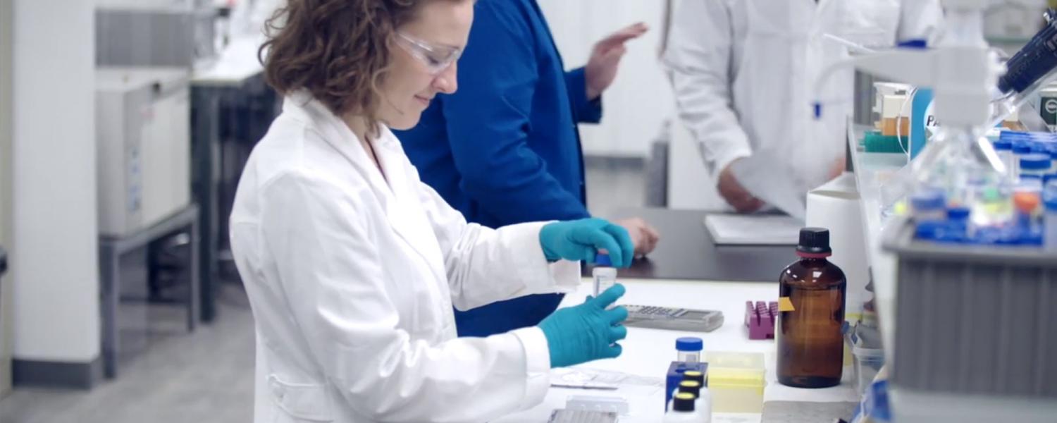
Step 2 – Visikol Labels and Scans Slides
Slides are labeled and scanned using Leica Versa Micrscopes. Alternatively, Visikol can also conduct confocal slide scanning, as well as more advanced imaging such as Imaging Mass Cytometry (IMC) and confocal microscopy.
Step 3 – Cloud Based Sharing, Viewing and Manipulating on BitSlide™
Visikol has developed an intuitive and easy-to-use cloud-based platform for viewing, downloading, and sharing large amounts of slide scanning data which is called BitSlide™. The platform allows researchers to easily collaborate, annotate, and manipulate their data and it is compatible with multiplex fluorescent data sets. The platform runs in an encrypted AWS environment within a web browser which means that clients do not need to download huge amounts of data and have specialized software just to view their images. Alternatively, Visikol can also ship hard drives of data to clients.
Step 4 – Image Analysis
Once the data for a project is completed, Visikol can execute image analysis for a client to transform the large amounts of data generated into actionable and quantitative insights. Visikol has a broad range of image analysis tools for many applications such as cell counting, digital pathology machine learning, immune cell population analysis, spatial analysis and blood vessel segmentation.
Affordable pricing and rapid turn-around time
Our standard slide scanning services are conducted with a 14-day turn-around time. However, we can deliver your results back to you in as little as 3 days depending on the size of a project. For our fluorescent imaging, we can image up to four channels simultaneously.
- Whole slide conversion to digital format.
- High resolution images up to 40x preserved digitally.
- H&E, IHC, and fluorescent stains.
- Digital images are compatible with Aperio ImageScope, ImageJ (with Bioformats), and Halo.
- Raw images can be returned via hard drive or the cloud.
- Secure and simple access to files.