
Tissue clearing eliminates light-scattering elements, enabling visualization of molecular targets within organs. It has diverse applications in fields like neuroscience and cancer research. A recent study titled ‘Combining tissue clearing and Fluoro-Jade C labeling for neurotoxicity assessment‘ explores using tissue clearing for neurotoxicity assessment, focusing on Fluoro-Jade C (FJ-C) labeling. FJ-C, a fluorescent tracer for neurodegeneration, is tested in cleared brain samples using various tissue-clearing methods. The research aimed to determine if FJ-C remains effective, and if combining it with tissue clearing can enhance neurotoxicity evaluation in animal models and human brain samples.
Various tissue-clearing techniques were employed in the study, each with distinct methodologies and procedures. These included the BABB method, wherein tissue samples underwent ethanol dehydration before immersion in a benzyl alcohol and benzyl benzoate solution. The CUBIC technique involved sequential immersion in diluted and concentrated CUBIC-L solutions, along with subsequent incubation in CUBIC-R solution. Moreover, for the SeeDB2 approach, saponin solutions and SeeDB2G solutions were used to facilitate clearing. Additionally, the Visikol-HISTO method employed methanol solutions followed by immersion in Visikol-HISTO-1 and Visikol-HISTO-2 solutions.
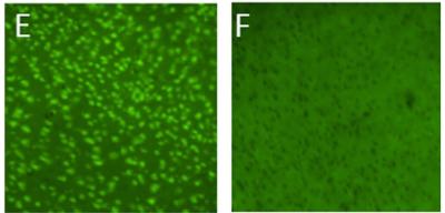
FJ-C labeling in tissue samples of KA- and saline-treated animals following incubation with FJ-C compatible tissue-clearing media: Visikol-HISTO (E and F). Left and right panels represent KA- and saline-treatment, respectively. Scale bar indicates 50 µm for all panels.
This study marks the first successful integration of tissue-clearing methods with Fluoro-Jade C (FJ-C) labeling, showcasing its compatibility for the detection of degenerating neurons. While tissue clearing has been employed for fluorescence imaging of molecular targets, the combination with FJ-C labeling was previously unreported. Results demonstrated that FJ-C remained specific for identifying degenerating neurons in Kanic acid (KA)-treated tissues, even after tissue clearing, suggesting the potential for broader application in various neurotoxicity models. Notably, compatibility varied among tissue-clearing methods, with FJ-C signal preservation observed in organic solvent-based techniques (BABB, uDISCO, Visikol-HISTO) and certain aqueous-based methods (SeeDB2, CUBIC-L). The successful amalgamation of these techniques paves the way for expanded three-dimensional images and holds promise for assessing neurotoxicity in entire cleared brains through light-sheet microscopy.
Visikol offers a wide range of tissue clearing reagents for transparentizing biological tissues and cell models, as well as advanced drug discovery solutions such as 3D cell culture assays and tissue imaging high content screening, AI solutions for histological analysis of tissue sections, and much more. To learn more about Visikol’s services, please contact us.