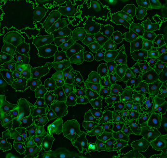
In the quest to unravel the intricacies of cellular biology, scientists have embarked on a journey into the microscopic realm, armed with powerful imaging techniques like Cell Painting. This high-content imaging method provides a detailed snapshot of cellular structures, but the real magic happens in the subsequent image analysis pipeline, with cell segmentation taking center stage. Cell segmentation is one of the first steps in cell painting analysis, which creates marked boundaries around the cells and helps in the precise measurement of insights into the cells painted with different dyes.

Figure 1. The above figure is a cell segmentation image created from a Cell Profiler pipeline: red lines outline the DAPI stained nuclei and green outlines the individual cell peripheries.
Cell Painting: A Palette of Fluorescence
Cell Painting is an innovative imaging technique that employs a palette of fluorescent dyes to label various cellular components. These dyes provide a vivid snapshot of cellular morphology and function from the nucleus to the cell membrane. As researchers generate rich, multichannel images, the subsequent image analysis becomes the key to unlocking most of the information in these microscopic masterpieces.
The Image Analysis Pipeline: A Roadmap to Discovery
The image analysis pipeline in Cell Painting is a well-orchestrated symphony of computational steps, each contributing to extracting meaningful insights from the raw images. At the heart of this pipeline is cell segmentation. This process identifies and outlines individual cells within the images, paving the way for a deeper understanding of cellular structures, textures, and behaviors. The image analysis team at Visikol has successfully developed this cell segmentation pipeline with a deeper understanding, which can be used for all cell types.
The Navigating Steps for Cell Segmentation:
- Preprocessing of Images:
Before diving into segmentation, raw images often undergo preprocessing. This step involves background subtraction, noise reduction, and normalization, ensuring the images are primed for accurate analysis.
- Nuclear Identification:
The journey of cell segmentation often begins with the identification of nuclei. Stains such as DAPI or Hoechst help highlight these cellular command centers, enabling segmentation algorithms to pinpoint the individual nuclei.
- Defining Boundaries:
Once nuclei are identified, the next challenge is outlining the boundaries of each cell. Fluorescent dyes targeting the cell membrane are crucial in defining the perimeters and completing the segmentation puzzle.
Some challenges of cell segmentation are the heterogeneity of cell populations as they are diverse in size, shape, and intensity, requiring robust segmentation algorithms. One of the big obstacles is cell-cell overlap, and advanced algorithms, including machine learning approaches, are employed to distinguish between overlapping cells. Image artifacts like uneven illumination or staining irregularities can complicate segmentation as well. Effective preprocessing techniques play a crucial role in mitigating the impact of these anomalies. Scientists at Visikol find methods to reduce these challenges and develop improved versions of the pipelines. They create various computational tools and techniques that aid cell segmentation.
These techniques are:
- Thresholding Techniques:
Basic thresholding methods assign pixel values above a certain intensity threshold to segmented objects. While straightforward, this approach can help in dealing with image heterogeneity. - Watershed Transformation:
The watershed transformation is a more sophisticated technique that mimics the flow of water to separate adjacent cells. It excels in handling overlapping cells and intricate cellular landscapes. - Machine Learning Algorithms:
Advanced machine learning algorithms, including deep learning approaches like convolutional neural networks (CNNs), have demonstrated remarkable success in cell segmentation. These algorithms learn intricate patterns from large datasets, making them powerful tools in this context.
The Cell Painting image analysis pipeline often benefits from open-source software tools and platforms. At Visikol, we use platforms such as Cell Profiler and ImageJ, which provide researchers with a user-friendly environment to implement and customize their segmentation workflows.
Significance:
Cell segmentation ensures precise identification and delineation of individual cells within the images captured through Cell Painting, which is fundamental for accurate phenotypic analysis, allowing researchers to explore subtle changes in cell morphology that may hold the key to understanding cellular responses to various stimuli. Cell segmentation goes beyond visual representation, enabling the extraction of quantitative morphological data. By quantifying cellular features such as size, shape, and subcellular distribution, researchers gain a detailed profile of cellular morphology. This quantitative approach enhances the robustness of analyses and facilitates comparisons across different experimental conditions. It extends to identifying and localizing subcellular structures within Cell Painting images. Understanding the spatial organization of cellular components provides insights into the dynamic interplay between different organelles. This is particularly valuable for unraveling complex cellular processes and their implications for cellular function. Thus, one of the components of cell painting, cell segmentation, creates a pathway for further analysis in the process of cell painting.
The significance of cell segmentation in Cell Painting cannot be overstated. It serves as the cornerstone for extracting valuable information from complex images, transforming them into a canvas that reveals the intricate details of cellular life. Image analysts at Visikol always strive to evolve the cell segmentation pipeline based on the end goals. Through precise delineation, quantitative analysis, and integration with molecular data, cell segmentation enables researchers to explore the hidden tapestry of cellular biology, opening new avenues for discovery and advancing our understanding of the complexities within each microscopic cell.
Future Vistas:
As technology continues its relentless march forward, the future of cell segmentation in Cell Painting holds promises of even greater accuracy, efficiency, and accessibility. The integration of artificial intelligence, advancements in machine learning algorithms, and improved computational resources are set to elevate cell segmentation to new heights. Reach out today to discuss your project with a member of our team!
