While the pathological hallmarks of Alzheimer’s disease (AD), a neurodegenerative disorder, have been established, there is ongoing research into additional biological mechanisms involved in the disease process. These mechanisms include calcium dysregulation and oxidative stress, where the secosteroid hormone Vitamin D (VitD) may play a role in protecting neurons against damage induced by reactive oxygen species (ROS) and other protective mechanisms. Studies have provided mixed results regarding the impact of VitD on AD pathology, with some suggesting positive effects and others indicating negative influences.

The Evaluation of VitD Deficiency in Chronic and Severe Alzheimer’s Disease
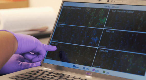
In a study conducted by Wong et al., the evaluation of VitD deficiency in chronic and severe AD utilized H magnetic resonance spectroscopy (), a common technique for monitoring metabolite changes in individuals with AD. Metabolite levels were measured in VitD-deficient adult transgenic AD mouse models, and assessments of ventricle volume and spatial memory performance were carried out. Behavioral studies were complemented by histological analyses, where brain samples from mice on a VitD-deficient diet and control diet mice were extracted, fixed, and scanned. Digital pathology image analysis tools developed by Visikol facilitated the evaluation of colocalization of regions of interest (ROIs) corresponding to the cortex and hippocampus with segmented astrocytes and amyloid plaques, allowing for the determination of the total number of astrocytes and amyloid plaques for each stained slice.
Summary of Results
The results revealed lower levels of N-acetyl aspartate and creatine in VitD-deficient mice compared to the control group at 12 months. Ventricle volume remained unchanged due to VitD deficiency. Lactate levels increased over time in VitD-deficient mice, and these increases were negatively correlated with changes in the primary latency to the target hole in the Barnes Maze. Surprisingly, VitD-deficient mice exhibited improved spatial memory performance compared to control diet mice. Additionally, VitD-deficient mice had higher astrocyte numbers in the cortex and hippocampus at 15 months. The study suggests that severe Vitamin D deficiency in human amyloid precursor protein and exon-9 deleted mutation of the human presenilin-1 protein APP/PS1 mice may prompt compensatory changes in metabolite and astrocyte levels, contributing to enhanced spatial memory performance. Further studies are needed to determine the impact of VitD deficiency and supplementation in patients with mild AD and in more aggressive models of AD.
Visikol offers a wide range of tissue clearing reagents for transparentizing biological tissues and cell models, as well as advanced drug discovery solutions such as 3D cell culture assays and tissue imaging high content screening, AI solutions for histological analysis of tissue sections, and much more. To learn more about Visikol’s services, please contact us.
