Introduction
Understanding the spatiotemporal patterns of gene expression is critical to decode complex biological events like disease and development. Digitally reconstructing the spatial patterns of expression in intact tissue paves the way for an unbiased and complete review of gene expression. This aids in understanding the molecular origin of developmental defect or disease pathogenesis for medical diagnostics. Visikol now offers next generation RNA and protein spatial profiling services powered by HCR RNA-FISH and IHC reagents from Molecular Instruments. HCR reagents enable multiplexed quantitative mapping of RNA and/or protein molecules with subcellular resolution and are rapidly becoming fundamental tools for drug discovery in gene therapy/RNA therapeutics.

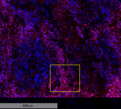
Figure 1. 4X imaging of B-actin RNA with HCR RNA-FISH (red) and DAPI (blue).

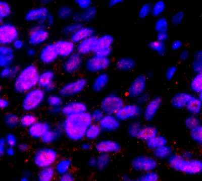
Figure 2. 40X imaging of B-actin RNA with HCR RNA-FISH (red) and DAPI (blue).
How it works
Background on Technology
The HCR imaging platform opens a new era for ISH and IHC, enabling multiplexed, quantitative, enzyme-free labeling of biomolecules in any sample type regardless of thickness or preparation. HCR RNA-FISH and HCR IHC allow simultaneous detection of up to 10 RNA and protein targets, respectively. Through this unique partnership, HCR RNA-FISH can be combined with Visikol’s wide array of in-house validated multiplex immunohistochemistry panels. This enables high-resolution co-detection of RNA and protein molecules with spatiotemporal and functional context in tissues. Furthermore, the technique is compatible with thick whole mount tissues in tandem with tissue clearing such that RNA can be imaged in large intact tissues using confocal or light sheet microscopy.

