
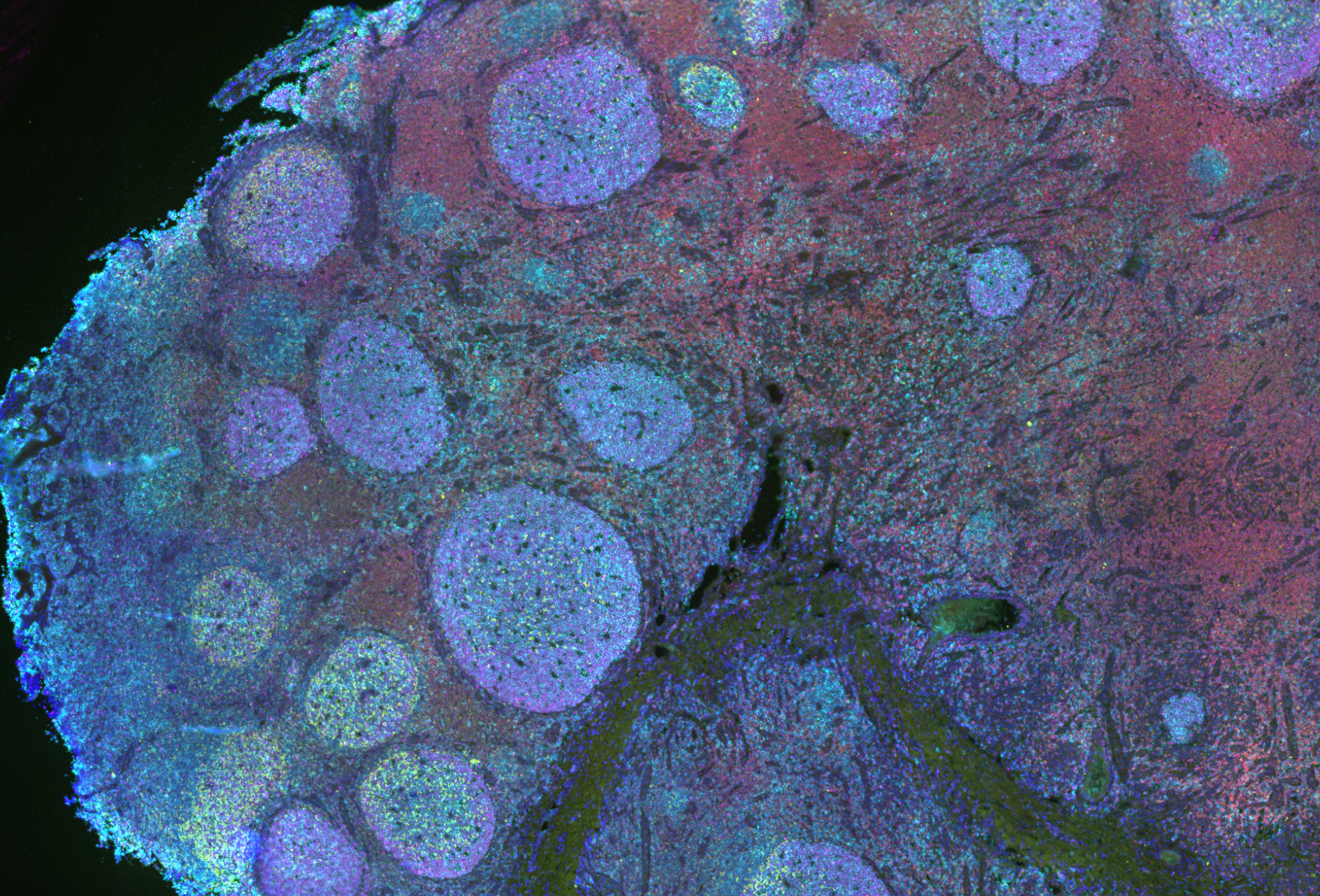
In the 21st century, modern medicine has become more and more complex with an ever-growing need for researchers to understand the body’s response to different disease states and medicinal treatment. One of the many tools that researchers can use to visualize cell populations, protein distribution, and treatment biomarkers is multiplex immunofluorescence which allows for many markers to be visualized simultaneously on a single sample. Visikol has developed a proprietary multiplex slide imaging approach which allows for the visualization of over ten markers per slide by conducting sequential rounds of 3-5 marker labeling and imaging. Complex labeling, such as multiple rounds of immunofluorescence, comes with a thorough optimization and validation for each antibody and panel prior to use in Visikol’s platform. Visikol’s antibody validation comes in two tiers: Tier 1, which is the standard validation and optimization for all projects and Tier 2, for project specific validation.
Tier 1 Antibody Labeling Validation
For each client project and when a new panel is developed, the Tier 1 standard validation and optimization is executed to ensure optimal and clean staining, which improves the analysis and image quality for each labeling. Upon receiving an antibody which has not previously been validated or comes from a different lot, a Visikol scientist will complete a concentration curve on tissue that is known to express the marker of interest. Utilizing the data sheet or previous publications, a range of 3-4 concentration dilutions is generated and labeled individually. After the completion of imaging, the optimal concentration is determined by examining label specificity, quality, and lack of background. Panels are performed on protein expressing tissues once a concentration is determined to confirm the labeling is successful with no crossover and minimal background. With these evaluations and every assessment that is done at Visikol, positive controls and secondary controls are the two key controls utilized with every experiment. Positive controls, which are tissues confirmed to express the respective markers, are utilized to confirm the staining in samples is in fact true and specific. The accompanying secondary control entails an experimental section to be labeled solely with the secondary antibodies and no primary, then imaged at the same intensity and magnification to ensure that the secondary antibodies, themselves, are not interacting with the tissue of interest giving false positives and high levels of background. Once each antibody has been validated and optimized labeling is completed on experimental samples with validation of antibody stripping following treatment.
Tier 2 Antibody Labeling Validation
Depending on the project or client needs, a multiplex design can require a second level of validation to generate optimal labeling and analysis alongside Tier 1 validation in which Visikol also offers an in-depth Tier 2 validation to clients as well. This validation includes the use of known negative control (ex. Knock-out mouse tissue or knock-out cell line) or an isotype control (ex. primary antibody lacking target specificity). Utilizing one of these two controls acts to determine that the antibody of choice is specific and is not cross-reactive with other markers, as no staining is expected in these two validation steps. For clients with personally generated antibodies or who show concerns of specificity with polyclonal antibodies, Visikol offers a competition assay control. This control allows the antibodies to interact with the specific antigen they were raised against prior to staining to ensure that no staining will be seen within the tissue, and therefor no cross-reactivity and specificity for their intended antigen.
These tiers of validation and optimization can be key when developing a multiplex panel to ensure the labeling and analysis is as true and efficient as possible. If you’d like to learn more, visit our Multiplex Technology Page, our Overview Page, or our recent blog post about the Clinical Applications of Multiplex Imaging.
And of course, please check out our new webpage detailing each of the panels in more detail and stay tuned for more panels coming in the near future!