Many human diseases are caused by morphological, physiological or genetic changes in cells. The challenges of disease diagnosis include the evolving nature of the disease, disease symptoms and the diagnostic process. Statistics on the frequency of missed diagnoses differ according to the symptoms or the eventual diagnosis. For hundreds of years, microscopy has been used to study the ultra-structural and physiological changes of the living cells. Based on the recent advancements in the field of microscopy, scientists have developed the cell painting technique, which is a high-content, multiplexed image-based assay used for cytological profiling. Since several compartments of cells are “painted” using different stains, cell painting has become a powerful phenotypic assay adopted by the academic screening centers and pharmaceutical industry for pathological diagnosis of cells as well as drug discovery.
What is Cell Painting?
Briefly, in cell painting, compound-induced phenotypic features will be imaged. With the help of high content microscopic imaging, phenotypic examination of physiological, metabolic or epigenetic perturbations in different cellular compartments can be visualized. Using different fluorescent dyes, various cellular compartments are stained and imaged. Finally, the image analysis workflow involves image segmentation and extraction of hundreds to thousands of parameters. One of the advantages of a cell-painting driven screen is that the phenotypic clustering will be independent of a known target. This enables identification of novel compounds with different mechanisms of action.
The team at Visikol is making great strides in the pathological diagnosis of cells thereby leveraging drug discovery efforts. If you are interested in utilizing this cell painting approach for your drug discovery projects, please reach out to our team to discuss your project. We are always interested to work together with our clients to develop customized drug discovery solutions to answer your research questions.

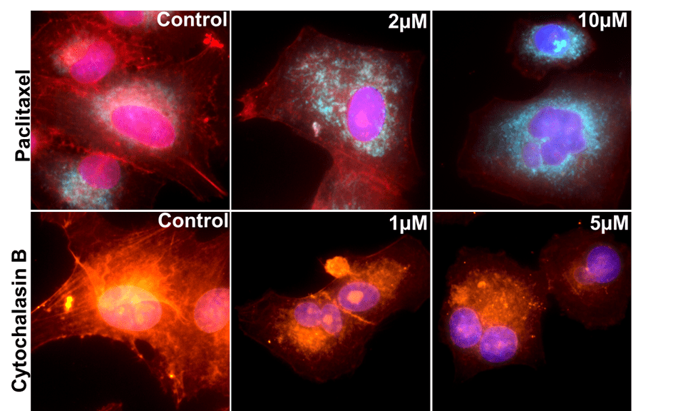
Top Panel: A549 cells treated with different concentrations of an anti-proliferative compound. Merged images show nucleus (stained by Hoechst), cytoskeleton (stained by Phalloidin), and mitochondria (stained by MitoTracker). The anti-proliferative compound inhibits cytoskeleton formation and induces disintegration of mitochondria.
Bottom Panel: A549 cells treated with different concentrations of Cytochalasin B. Merged images show nucleus (stained by Hoechst), cytoskeleton (stained by Phalloidin), and nucleoli (stained by SYTO14). Cytochalasin B inhibits cytoskeleton formation and disintegrates nucleoli.
