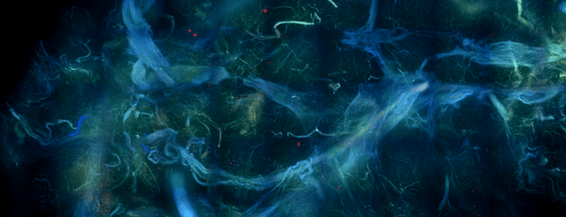
Confocal z-projection of a melanoma sentinel lymph node biopsy
At Visikol, our scientists are skilled in various types of microscopy that are useful in the pharmaceutical industry for many applications, including drug discovery and development and quality control. One of the most common types of microscopy is light microscopy; this technique uses visible light to observe samples and allow the visualization of tissue structures. Light microscopy is a critical tool for the diagnosis of many diseases, including cancers, infections, and inflammatory disorders.
The Importance of Microscopy
In drug development, microscopy is important because it can be used to study the effects of potential drugs on tissues, for example it is used to study the localization and expression of proteins in conjunction with fluorescent labeling techniques to visualize specific structures within tissues. Fluorescence light microscopy is one of the most widely used tools in pharmaceutical research. Fluorescent labeling is done by first blocking the sample with a blocking buffer in order to prevent nonspecific binding of the primary antibody. Next, a primary antibody is used to bind to a specific antigen of interest. After the sample is incubated in a primary antibody, it is labeled with a fluorescent dye conjugated secondary antibody to bind to the primary antibody. The secondary antibody will allow the sample to be visualized with fluorescent microscopy. There are many kinds of light microscopy, but two popular types of fluorescent light microscopy are widefield and confocal microscopy.
Widefield Microscopy
Widefield microscopy is a type of microscopy that uses a light to show the whole sample. This technique is beneficial because it is highly sensitive, allowing for the detection of low levels of fluorescence in samples. There is low photodamage because widefield microscopy does not use a high intensity laser light, therefore samples can be imaged multiple times if needed with minimal damage. Thinner samples are best imaged using widefield microscopy. One limitation of widefield microscopy is that it may not provide high resolution images of the tissue; to get higher resolution and more detailed images, confocal microscopy can be used.
Confocal Microscopy
Confocal microscopy is beneficial because it uses a pinhole to reject out-of-focus light, which can reduce background noise and improve contrast in images; this makes it easier to visualize subtle details within tissues and allows for thicker samples to be imaged compared to when imaging with widefield microscopy. Confocal microscopy also allows for the visualization of samples with high spatial resolution and can obtain 3D images of the sample, which is especially useful for studying tissues and cells in great detail. Due to the high quality images produced, confocal microscopy may take longer than widefield microscopy.
Overall, Visikol provides different microscopy services depending on our Client’s needs. Contact us to work with our expert team of scientists.
