What is Immunohistochemistry? What is immunofluorescence? What separates these techniques, and when is the best time to choose one over the other? These terms are frequently used interchangeably, which can lead to confusion when preparing for a tissue or cell labeling microscopy experiment. This blog will unravel these methods, discuss their pros and cons, and help aid in deciding which microscopy technique is the best for your next experiment.
Immunohistochemistry and Immunofluorescence are the main techniques used in histology. Both assays involve the use of antibodies to detect protein while maintaining cellular structure. Each of these techniques also require the use of a microscopes to visualize the structures of the labeling or staining that has been performed.

Immunofluorescence

Immunohistochemistry
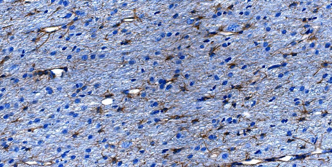
Immunohistochemistry (IHC) is the most commonly used term when discussing microscopy techniques performed on tissue samples, both paraffin embedded and frozen. IHC uses antibodies to detect specific proteins, however, it is visualized using a colored chromogen and counterstain (e.g. hematoxylin, Methyl Green or Nuclear Fast Red). Antibodies bound to enzymes, most commonly horseradish peroxidase (HRP) or alkaline phosphatase (AP) are used with various chromogens (e.g. AEC, TMB, StayYellow and DAB) to generate an array of colors. IHC is then visualized using a brightfield microscope.
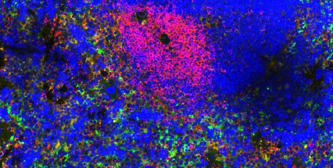
While similar, IF relies on antibodies attached to a fluorophore or fluorescent dye (e.g. FITC, Texas Red, Cy5 and Alexa Fluors) in order to be visualized. This method is used in combination with a nuclear label, most commonly DAPI or Hoechst, and is visualized with a fluorescent microscope. The primary antibody will bind to the target proteins and through direct or indirect detection, the fluorescent microscope will visualize the antibodies using lasers operated at specific wavelengths.
While both tissue labeling techniques can be utilized when detecting markers of interest, each has their pros and cons to consider. When labeling tissue for distinct cell populations with prevalent tissue architecture and long-term storage, IHC is commonly the preferred method. However, when interested in co-localization of markers that are not commonly prevalent for IHC, IF methods combined with multiplex techniques is the preferred method to obtain the most amount of data from a single specimen. Each technique while having its own pros and cons can give a researcher a deep insight into the tissue environment.
| Pro | Con | |
|---|---|---|
| Immunohistochemistry (IHC) |
|
|
| Immunofluorescence (IF) |
|
|
Visikol and its cutting-edge Multiplex technology utilize the IF techniques and standard IHC methods which offer a new paradigm in research, opening a whole new door to understanding the tissue microenvironment, disease progression, and treatment. If you’d like to learn more, visit our Multiplex Technology Page, our Overview Page, or our recent blog post about the Clinical Applications of Multiplex Imaging and how it can be utilized as a Clinical Tool, or reach out to a member of our team.
