
 Just about anyone who has attempted to fluorescently label whole tissue using antibodies has eventually realized that there are a million and one factors that go into the quality of the finished product. Finding a primary antibody that successfully sticks to your target and pairing it with an appropriate fluorophore is just the beginning of the process. Optimization is often necessary to correct for any number of “imperfections” in the images one acquires.
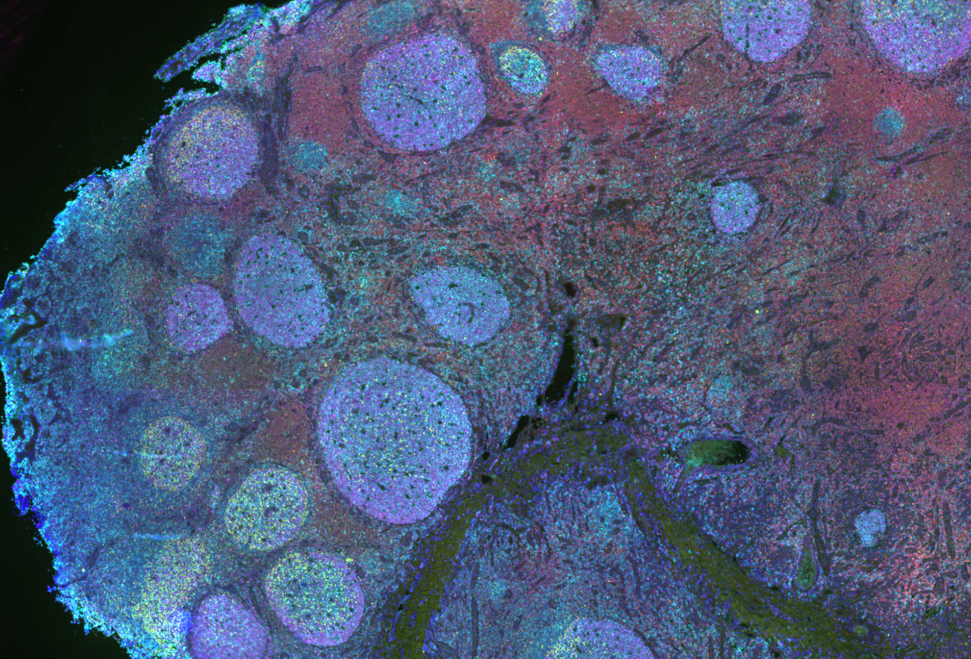
One area where these imperfections easily arise is in the uniform labeling of an epitope throughout the tissue.
Here are some items to keep in mind when determining the ideal protocol for your labeling needs:
- It takes time for antibodies to move throughout a tissue. The only way to ensure your labels can be applied evenly is to give them time to reach all targets. Pieces of tissue as small as 1 mm may only require 5-10 hours of antibody incubation, but times ramp up quickly as you increase the size. As a general rule, for each doubling of the thickness of the tissue, you’ll want to triple the antibody incubation time. As an example, one hemisphere of a mouse brain may be adequately labeled after 1.5 days of exposure to antibody, but making the jump to a whole mouse brain can increase the ideal incubation time to 5 days.
- Dense tissues can increase this time even more. Optimizing antigen retrieval, penetration, and permeabilization steps is a whole topic unto itself, but it must be kept in mind that antibodies depend on open spaces throughout the tissue in order to move to the epitope. If your tissue isn’t very porous, or is perhaps resistant to the steps that increase the ability of the antibody to flow among cells, you may need to increase incubation time even further to allow for adequate coverage.
- More antibody isn’t always better. While it may seem that flooding tissue with an antibody is the surest way to cover it from top to bottom, that is certainly not always the case. Antibodies are often fairly large relative to the spaces they have to work with. If there are too many of them binding quickly and readily near the surface, they may block the path for the rest to move farther into the tissue. When optimizing antibody concentrations, make sure to image all the way through the tissue when possible. You may find that, at times, less is more!
For more details on immunolabeling, along with many other aspects of tissue imaging in your experiments, be sure to check out Visikol’s online resources, including our Tissue Clearing Ebook. Visikol’s scientists are also ready to apply their experience to the imaging needs of your research! Contact us today for more information.