In 2022, Visikol launched a new imaging service in partnership with Standard BioTools Inc, this partnership has allowed us to bolster our imaging capabilities with the support of an industry leader in imaging. With new imaging services, our Business Development team is frequently asked about the use case of each imaging modality. The following few paragraphs will delineate the different imaging modalities and the use cases for each.
The process for designing a study plan requires consideration into one’s desired endpoints. Endpoints can be pathology review, discovery data, or clinical insights. To get to each endpoint, there is a different modality.
Standard Imaging
In the case of standard imaging, H&E, DAB, Masson’s Trichrome (and others) are staples of staining and brightfield imaging. These stains are often easy to use, efficient and cost effective. The use case is for projects of scale, which require simple area defining analysis or pathology review. Brightfield projects also are typically fast turnaround, making it a great fit for a library of samples that need digitization.


Multiplex Immunofluorescent
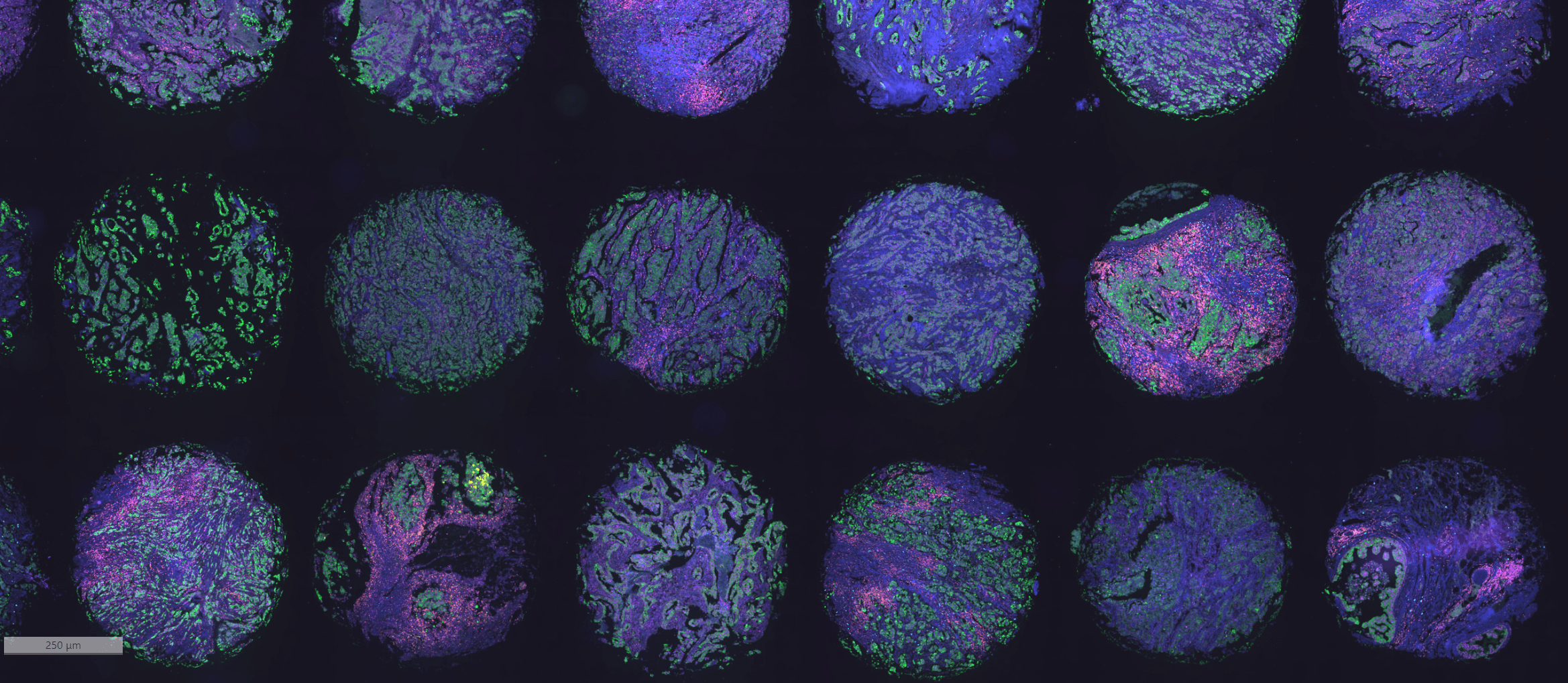
The next imaging method is IF (immunofluorescent) – specifically multiplexing. Multiplex IF refers to the labeling of a tissue section with more than 2 antibodies and fluorescently imaging antibodies to create an image. These projects are often more labor intense, and expensive than standard imaging. The pricing and turnaround time for these projects are based on complexity. In other words, a sample imaged with 3 antibodies or 20 antibodies will have a major impact on materials needed and effort required to produce images. The use case for IF is for preclinical or clinical tissues with a medium to high throughput sample quantity. IF can prove useful in drug discovery research as well as translational research and is a great starting point for understanding the TME (tumor microenvironment).
Imaging Mass Cytometry (IMC)
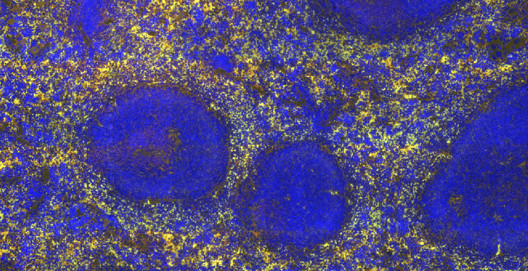
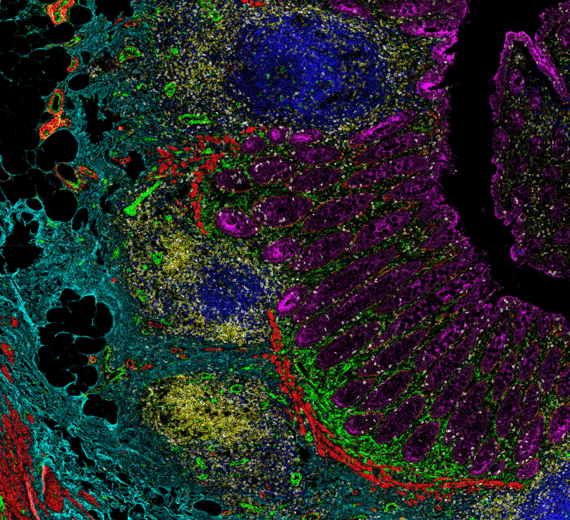
Similar to IF imaging, is IMC (Imaging Mass Cytometry). At Visikol, IMC imaging is available through the Hyperion+ Imaging System and provides researchers the opportunity to generate very large amounts of data from small sections of tissue. Unlike IF, IMC allows researchers to choose up to 40 markers per tissue and there is no background noise because the process is a metal-binding labeling approach. While IMC is more expensive than IF, it provides a greater amount of data that is best used for clinical samples in which researchers need to derive as much data as possible out of valuable tissue samples.

At Visikol, we help our clients with all three of the aforementioned imaging approaches. Our team has worked on projects of all types and are happy to help with you about your next project!
