
Fluorescence in situ hybridization (FISH) is a molecular technique used to detect and localize specific DNA or RNA sequences within cells, tissues, or whole mount organisms. This technique is based on the use of fluorophore-labeled probes that hybridize complementary DNA or RNA target sequences, allowing for the visualization and identification of specific genetic loci or transcripts. FISH has become an essential tool for both research and diagnostic applications, allowing for the identification of genetic aberrations and disease-associated biomarkers in a variety of tissues and cell types.
FISH Tissue Labeling
The process of FISH tissue labeling with a fluorescent probe involves several key steps. The first step is the selection and design of the appropriate probe sequence. The probe is typically a short, single-stranded DNA or RNA molecule that is complementary to the target sequence of interest. For FISH, the probe is labeled with a fluorophore allowing for its detection and visualization under a fluorescence microscope. The selection of the probe sequence is critical, as it determines the specificity and sensitivity of the FISH assay.
Once the probe sequence has been selected, the next step is to prepare the tissue sample for labeling. This involves fixing the tissue in a solution of formaldehyde, paraformaldehyde, or other fixatives, which preserve the tissue morphology and prevent the degradation of the nucleic acids within the sample. The fixed tissue is then permeabilized with proteases to allow the probe to penetrate the cell membrane and hybridize to the target sequence. Protease treatment also increases hybridization efficiency and reduces background fluorescence of the sample. In the case of whole mount FISH, the organism may need to be cleared, or made transparent, prior to fixation by using clearing agents meant for large tissues, such as Visikol® HISTO™-1 or HISTO™-2. In addition to these two reagents, Visikol® HISTO™-M can be utilized for cell culture models and tissues less that 1mm thick.
After the tissue has been fixed and permeabilized, the next step is the hybridization of the fluorophore-labeled probe to the target sequence. The probe hybridizes to the complementary target sequence within the tissue, allowing for its detection and localization under a fluorescence microscope. After hybridization, the tissue sample is washed to remove any unbound or nonspecifically bound probes. This step is critical to minimize background fluorescence and increase the signal-to-noise ratio of the FISH assay. The washed tissue sample is then ready for imaging using a fluorescence microscope.
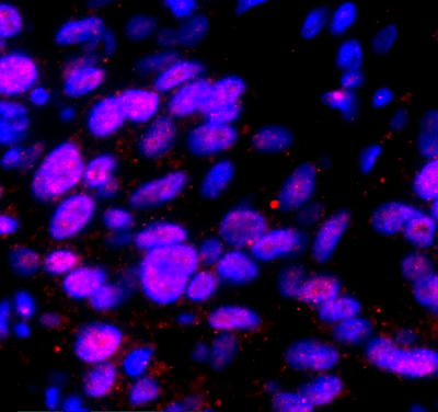
FISH and Immunofluorescence
Immunofluorescence imaging is a complementary technique that can be used in conjunction with FISH to visualize specific proteins or other cellular structures within the tissue sample. This technique involves the use of antibodies that are labeled with fluorophores, which recognize and bind to specific protein antigens within the tissue. The tissue sample is incubated with the labeled antibody, allowing for the specific detection and localization of the protein of interest. Both techniques can utilize multiple fluorophores to create multiplex labeling panels to visualize multiple target sequences at once. Here at Visikol, we specialize in highly multiplexed immunofluorescence imaging techniques involving 10+ different antibodies, made possible by our proprietary EasyPlex™ antibody stripping reagent and co-registration software.
Fluorescence in situ hybridization (FISH) is a powerful technique for the visualization and identification of specific genetic loci or transcripts within cells or tissues. The process of tissue labeling with a fluorescent probe involves several key steps, including the selection and design of the appropriate probe sequence, tissue fixation and permeabilization, probe hybridization, washing, and imaging. Immunofluorescence imaging can be used in conjunction with FISH to visualize specific proteins or other cellular structures within the tissue. The combination of these techniques has a wide range of applications in both research and clinical settings, allowing for the identification of disease-associated biomarkers and rapid diagnosis and treatment.
For more information on Visikol’s HISTO™ clearing reagents or our multiplex immunohistochemistry services, please reach out to a member of our team.
References
Egger D, Troxler M, Bienz K. 1994. Light and electron microscopic in situ hybridization: non-radioactive labeling and detection, double hybridization, and combined hybridization–immunocytochemistry. J Histochem Cytochem 42:815–822.
Jin L, Lloyd RV. 1997. In situ hybridization: methods and applications. J Clin Lab Anal 11:2–9.
Rautenstraub BW, Liehr T. 2002. FISH Technology. Berlin: Springer-Verlag.